Essential Question: What are the structures found in human skin and how do they function?
Introduction: The integumentary system includes the skin, hair, nails, sebaceous glands, and sweat glands. These organs provide a protective covering for deeper tissues, aid in regulating body temperature, reduce water loss, house sensory receptors, synthesize various chemicals, and excrete small quantities of wastes.
Directions: Use your notes and the textbook to complete the following activities.
NOTE: Cut and Pasted answers will receive no credit!
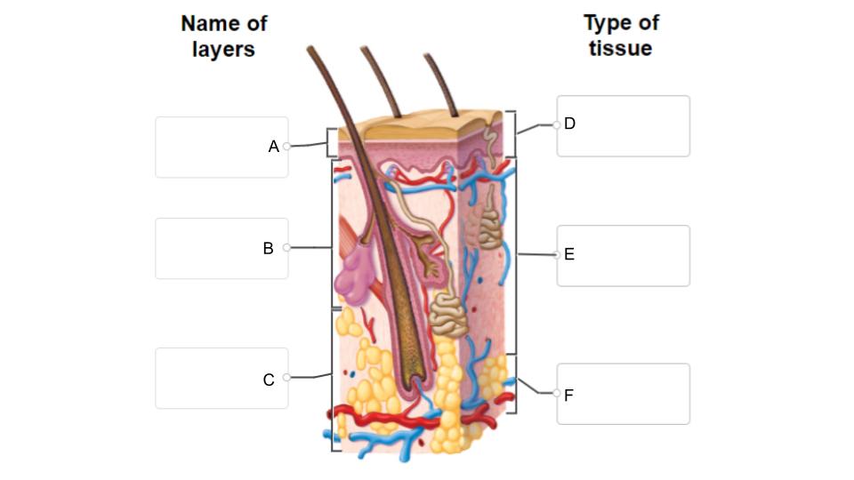
Match the layers of skin and the type of tissue to the correct location on the diagram.
| Draggable item | arrow_right_alt | Corresponding Item |
|---|---|---|
Dense Fibrous Irregular Connective Tissue | arrow_right_alt | A |
Hypodermis (Subcutaneous) | arrow_right_alt | B |
Stratified Squamous Epidermis, keratinized | arrow_right_alt | C |
Epidermis | arrow_right_alt | D |
Areolar and Adipose | arrow_right_alt | E |
Dermis | arrow_right_alt | F |
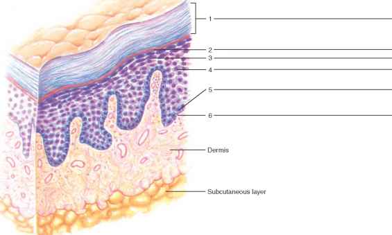
Match the layer of the epidermis to its location on the image
| Draggable item | arrow_right_alt | Corresponding Item |
|---|---|---|
Stratum lucidum | arrow_right_alt | 1 |
Stratum spinosum | arrow_right_alt | 2 |
Stratum germinativum (basale) | arrow_right_alt | 3 |
Stratum granulosum | arrow_right_alt | 4 |
Stratum corneum | arrow_right_alt | 6 |
Which layer of the skin is found only in thick skin and not in thin skin?
What are two other differences between thin and thick skin?
What are keratinocytes?
What are melanocytes? Why do we need them?
In which layer of the epidermis are melanocytes found?
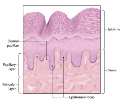
Distinguish between the two layers of the dermis
What structure forms the dermal ridges?
Based on the types of tissue found in the subcutaneous (hypodermis) layer, what do you think is its primary purpose?
Upload the labeled drawing of the scalp slide you made after completing this lab station.
The materials for this section are on the gray cart.
Go to Lab Table #3 and follow the instructions in order to complete this section.
Human skin color comes from three different sources. In the absence of any other colorizing agents, human skin is pinkish in color, due to the presence of oxygenated blood flowing through the dermis and subcutaneous layers of the skin. Human skin color also comes from the presence of two different pigments in the skin. Carotene is a yellow pigment, stored in the epidermis, dermis, and subcutaneous layer. The presence of carotene gives the skin a yellowish coloration. Having a very thick stratum corneum will also impart a yellowish coloration to the skin (this trait is mostly restricted to people of East Asian ancestry). Melanin is a brown/black pigment and is the main agent of human skin coloration. The more melanin one has in their skin, the darker their skin is. Melanin is found in the epidermis, manufactured by cells called melanocytes, found in the stratum basale. The skin on the palm of your hand and on the sole of your foot tends to be devoid of melanin pigment, regardless of how dark the rest of your skin is.
Observe the color of the skin covering the palm of your hand. What color is it?
Of the three coloring agents of human skin, which one(s) are responsible for this color?
Using the provided photos, observe pigmented and unpigmented skin. Make a labeled drawing of what you observe. Potential labels include: epidermis, dermis, pigment, stratum basale (germinativum,) stratum corneum, etc. These labels should be present on BOTH drawings.
Upload a photo of the drawing on your paper.
What SIMILARITIES did you observe between the two samples of skin? Name at least 2
What DIFFERENCES did you observe between the two samples of skin? Name at least 2.
These materials are on table 4.
Go to Lab Table #4 and follow the instructions in order to complete both of these stations.
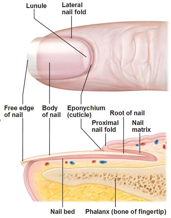
What are nails made of?
How are nails connected to the skin?
Using the magnifying glass, examine your fingernails and those of another person. Describe any differences that you see (color, striations, shape, etc.)
The instructions are in the packet with the materials for station #3.
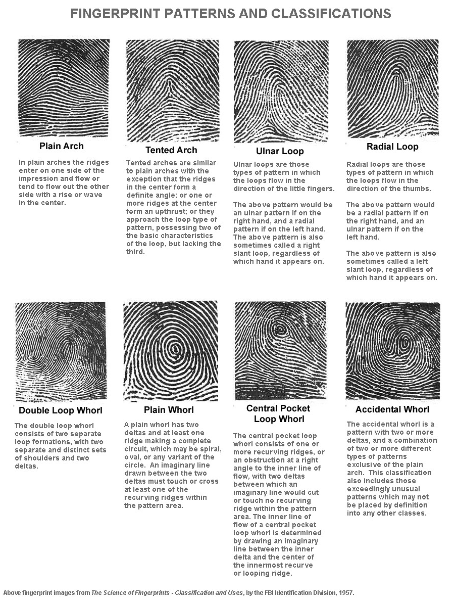
Follow the instructions to complete this station. You may use the reference at the link to help you complete this station. https://umbc.edu/stories/how-did-i-get-my-own-unique-set-of-fingerprints/
Describe the pattern you see on your thumb.
Do your other fingers have a similar pattern to your thumb? If no, explain.
Compare the patterns on your fingers and another person. Are they similar or different? Explain.
What anatomical structures create your fingerprints?
Go to Table #5 and follow the instructions in order to complete this station.
Fill in the table after following the instructions at this lab table.
How is the phenomenon of sensory adaptation helpful to humans?
Pain receptors do not adapt. Why do you think this is important and useful?
Observe the skin of your anterior and posterior forearm up close. If you have a magnifying glass, that may help your observations. How do the hairs differ in these two areas? List at least TWO differences.
Examining the hair root
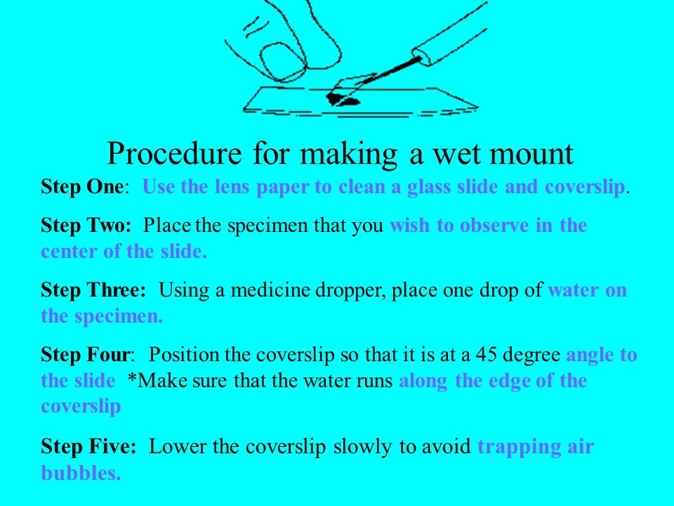
Make a wet mount slide of one of your hairs:
Pull out a single hair and place the root end on a microscope slide. (Hairs shorter than 1 inch work best.)
Place a drop of water on top of the hair then cover it with a cover slip. Try not to have any air bubbles around the hair.
Observe the root and shaft of the hair and note the scale-like parts that make up the shaft.
Sketch the hair at 100x magnification (low power.) AND Label the root and shaft in your drawing.
Tips for success:
Make sure that your sample includes the "root end."
If your cover slip seems to be floating, blot the top of your slide with a small piece of paper towel to absorb the excess water.
Upload the LABELED drawing of what you see under the microscope. Include the total magnification.
Which tested area had the most sweat glands?
Present a hypothesis to explain this difference.
Match the description with the correct structure or function.
| Draggable item | arrow_right_alt | Corresponding Item |
|---|---|---|
Dermis | arrow_right_alt | Becomes active during puberty |
Subcutaneous layer (Hypodermis) | arrow_right_alt | Causes hair to stand on end and goose bumps to appear |
Sebum | arrow_right_alt | Inner layer of skin |
Hair follicle | arrow_right_alt | Responds to elevated body temperature |
Keratin | arrow_right_alt | General name of entire superficial layer of the skin |
Epidermis | arrow_right_alt | Tube-like part that contains the root of the hair |
Melanin | arrow_right_alt | Hard protein of the hair and nails. Also found in keratinocytes |
Arrector pili muscle | arrow_right_alt | Epidermal pigment |
Eccrine sweat gland | arrow_right_alt | Pigment-producing cell |
Melanocyte | arrow_right_alt | Gland that secretes an oily substance |
Sebaceous gland | arrow_right_alt | Oily secretion that helps to waterproof body surfaces |
Apocrine sweat gland | arrow_right_alt | Binds skin to underlying organs |
Match the structure to its location
sebaceous glands
melanocytes
sweat gland
arrector pili muscles
blood vessels
hair root
adipose tissue
epidermis
dermis
hypodermis
Justify the following statement: Sebaceous glands are NOT a necessary component of thick (glabrous) skin.
The most superficial layers of the epidermis become less viable & ultimately die. What two factors account for this natural demise of the epidermal cells?
Name two processes involved in maintaining homeostasis that would be compromised if a person’s skin were to be replaced with scar tissue.