Copy and pasted answers will not be accepted.
Materials: Long bone sections, gloves, magnifying glass
***Videos are for reference if you have questions!
Examine the long bone sections and locate the periosteum on the fresh bone at this station. Describe the appearance of this structure.
Observe the external structure of the diaphysis and epiphysis on the bone. How does the structure of the bone differ in these areas? What types of other tissues or structures do you observe besides bones?
Locate an epiphysis and its articular cartilage. Rub the cartilage with your gloved hand and describe its texture.
What type of cartilage forms the articular cartilage?
What is/are the purpose(s) of this type of cartilage?
Observe the internal bone structure of the diaphysis and epiphysis on the clean, dry bone. How does the structure of the bone differ in these areas?
Draw the section of long bone at your table. Label as many of the parts as you can identify, including, but not limited to, the diaphysis, epiphysis, spongy bone, compact bone, yellow marrow, red marrow, periosteum, epiphyseal plate and articular cartilage.
Match the description of the parts of a long bone with the correct term.
| Draggable item | arrow_right_alt | Corresponding Item |
|---|---|---|
Fibrous membrane that covers the surface of bone; has the potential to form bone during growth periods and fracture healing | arrow_right_alt | Diaphysis |
Thick layer of hyaline cartilage; provides basis for production of spongy bone; only places where long bones grow in length after birth | arrow_right_alt | Medullary cavity |
Covers the bone at joint surfaces of the epiphysis; provides slick surface that reduces friction and allows the joint to move freely | arrow_right_alt | Epiphysis |
Tubular shaft of a long bone; hollow cylinder with walls of compact bone | arrow_right_alt | Epiphyseal plate |
The ends of a long bone; consists of a thin layer of compact bone-filled with spongy bone | arrow_right_alt | Endosteum |
Thin membrane that lines the medullary cavity | arrow_right_alt | Periosteum |
Hollow cylinder found in the diaphysis; filled with yellow bone marrow | arrow_right_alt | Articular cartilage |
Obtain a prepared microscope slide of compact bone. Select an osteon and draw or photograph it under high power (400x.) Label the following structures: osteon, canaliculi, lacunae, and Haversian canal.
How many complete osteons are visible under 400x?
Match the terms with their description.
| Draggable item | arrow_right_alt | Corresponding Item |
|---|---|---|
small canals that connect the lacunae in compact bone | arrow_right_alt | osteon |
concentric layer that surround the central canal of an osteon | arrow_right_alt | canaliculi |
a descendant of an osteoblast that resides within the lacunae of compact bone | arrow_right_alt | Haversian canal |
central canal of an osteon that contain nerves and blood vessels | arrow_right_alt | lamellae |
cell-shaped empty spaces found in bone and cartilage | arrow_right_alt | osteocyte |
functional unit of compact bone | arrow_right_alt | lacunae |
Observe the following prepared microscope slides of cancellous (spongy) bone. Select a section and draw it under 400x. Label the following structures: Osteocytes, Red Marrow, Trabeculae.
Take a photo of your drawing and insert it in the space below.
Match the following terms to their description.
| Draggable item | arrow_right_alt | Corresponding Item |
|---|---|---|
Type of marrow mostly found in flat bones and the ends of adult long bones. COntains stem cells that differentiate to form blood components such as RBCs, WBCs, and platelets. | arrow_right_alt | osteoblasts |
Cell that secretes the matrix that forms bone | arrow_right_alt | osteoclasts |
Type of marrow used for fat storage. Also, contains stem cells for fat, cartilage and bone production. Can produce red blood cells in times of dire need. | arrow_right_alt | red marrow |
Cell that causes bone resorption and is important in bone remodeling | arrow_right_alt | trabeculae |
Lightweight, porous material that fills the interior spaces of most bones | arrow_right_alt | yellow marrow |
Materials: Labeled bones
Watch the video about bone shapes and then use the information to sort the bones at the station by shape.
Locate the long bones of the skeleton. Where are they located?
Are the areas of the skeleton containing long bones considered part of the appendicular or the axial skeleton?
Examine the wrists and ankles of the skeleton. What types of bones are
located in these areas?
Examine the bones of the skull. How are these bones classified?
Locate a sesamoid bone in this skeleton. Describe its appearance. Where is it located?
Observe the bones of the spine. Under what name are these bones collectively known?
What is the shape classification of the bones in the spine?
Organize the bones at this station into the major categories of bone shapes.
A
B
C
D
E
F
G
H
I
J
K
Long
Short
Flat
Irregular
What physical characteristics do all of the bones classified as irregular bones have in common?
Which bones present at this station would most likely be the site of MOST hematopoiesis in an adult?
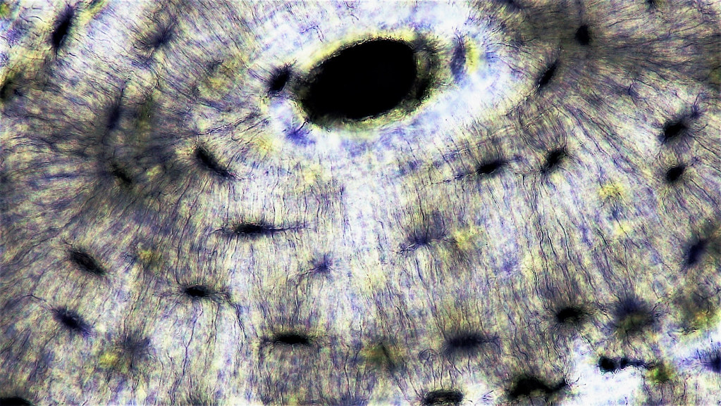
Examine the slide of hyaline cartilage at both low (100x) and high (400x) power magnifications.
Draw a section of hyaline cartilage under high power (400x) and label the perichondrium, chondrocytes and lacunae.
Take a photo of your drawing and insert it in the space below.
Where in the skeletal system would you find hyaline cartilage?
Based on the information in the videos, what is meant by the term ossification?
What are the two types of ossification?

Match the terms to the correct stage in the ossification process.
Growth continues at epiphyseal plate until full grown and plate fuses
Hyaline cartilage model
Blood vessels invade
Medullary cavity forms
Hyaline cartilage remains only in epiphyseal plate and articular surfaces
Cartilage calcifies in the center of the diaphysis
Secondary ossification center develops
Primary ossification center forms
A
B
C
D
E
There are two major categories of materials found in the matrix of bone: organic and inorganic.
Describe the inorganic substances found in the extracellular matrix of bone.
Describe the organic substances found in the extracellular matrix of bone.
In children suffering from rickets, the bones are so flexible they bow under the child’s weight. Which chemical component in the bony matrix is in short supply: collagen or minerals? Explain.
Osteocytes are surrounded by a dense, bony matrix. How do they get the nutrients they need to survive?
Explain, in detail, how weight-bearing exercise causes bones to increase in density and strength.
Watch the video about calcium homeostasis in blood and bone. Use the information in the video to help you correctly categorize the actions with the hormone responsible.
Increases calcium levels in body fluids
Decreases calcium levels in body fluids
Stores calcium in bone
Releases calcium from bone
Calcitonin
Parathyroid Hormone
What is meant by the term bone remodeling?