Upload a photo of your notes.
Purpose:
In this pre-lab you will:
Identify the parts of the microscope and their uses
Learn safe microscope operations
Prepare to use the microscope in lab next class period
Click the following link to go to the Virtual Microscope Lab. When you arrive, click “Launch Activity” to enter the laboratory. This virtual microscope is VERY similar to the actual microscope that you will be using in class next class period.
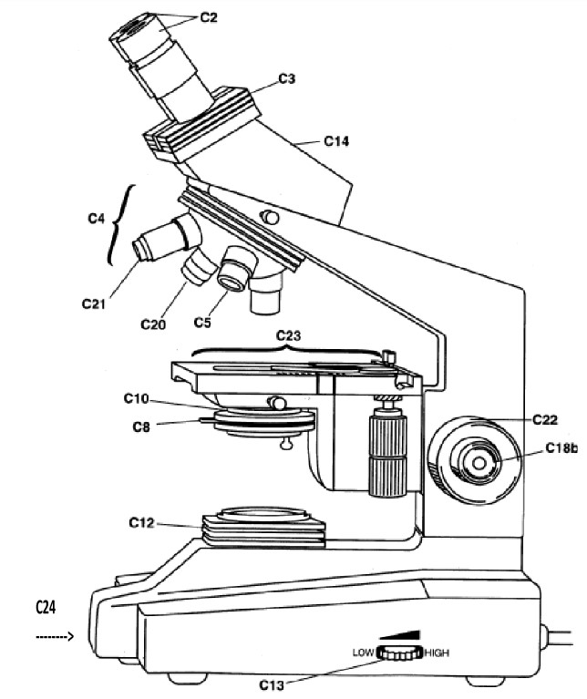
Match the part of the microscope with its location on the diagram
| Draggable item | arrow_right_alt | Corresponding Item |
|---|---|---|
Abbe condenser | arrow_right_alt | C24 |
diaphragm | arrow_right_alt | C12 |
rheostat (dimmer) | arrow_right_alt | C8 |
light source | arrow_right_alt | C10 |
mechanical stage | arrow_right_alt | C4 |
coarse adjustment knob | arrow_right_alt | C2 |
base | arrow_right_alt | C14 |
fine adjustment knob | arrow_right_alt | C13 |
head | arrow_right_alt | C23 |
ocular (eyepiece) | arrow_right_alt | C18b |
objective lens | arrow_right_alt | C22 |
Match the microscope structure with its function
| Draggable item | arrow_right_alt | Corresponding Item |
|---|---|---|
base | arrow_right_alt | Moves stage up and down in large increments (DO NOT use it on high power above 10x) |
mechanical stage | arrow_right_alt | Moves stage up and down in small increments. Use after coarse adjustment. (If using the 40x or 100x objectives it is the ONLY one you use) |
objective lenses | arrow_right_alt | Holds and positions the slide |
Abbe condenser | arrow_right_alt | Adjusts intensity of light |
coarse adjustment knob | arrow_right_alt | Controls amount of light that can reach specimen by changing diameter of opening |
fine adjustment knob | arrow_right_alt | Raises or lowers the diaphragm and thereby changes angle of light and intensity |
diaphragm | arrow_right_alt | The part you look through! Usually magnify 10x or 25x |
ocular (eyepiece) | arrow_right_alt | Magnifying lenses (4x, 10x, 40x, 100x (oil immersion lens) |
rheostat (dimmer) | arrow_right_alt | Lowest part of the microscope; supports the rest |
Part 2: Questions
Before you can begin observing specimens, you should familiarize yourself with the parts of the microscope and their functions. Click “Guide” at the bottom of the screen, then select “Overview”.
Link to Virtual Microscope
What is the proper way to carry a microscope?
What happens when the revolving nose-piece is rotated?
How can you determine the magnification of the ocular lens on your microscope?
Match the color of the objective lens with its magnification.
| Draggable item | arrow_right_alt | Corresponding Item |
|---|---|---|
yellow | arrow_right_alt | 40x (high power) |
white | arrow_right_alt | 10x (low power) |
blue | arrow_right_alt | 4x (scanning) |
red | arrow_right_alt | 100x (oil immersion) |
If the ocular lens has a magnifying power of 25X, and the low power objective lens is being used, what is the total magnification of the microscope? Show your work!
Now click “Microscope Care”.
Why must you never touch the lens of a microscope with your fingers?
Under what circumstances would immersion oil be used?
If you need to move the stage to the left, which part would be used?
What is the difference between the coarse focus (adjustment knob) and the fine focus?
The ocular lens of your microscope has several smudges. What would you use to clean the lens: paper towels or lens paper? Why?
Now click “Next”.
If you were trying to simply locate a grouping of cells under the microscope, which lens would be used? Explain using the term field of view.
Use Chapter 3 in the Online Textbook to help you define the following terms:
| Draggable item | arrow_right_alt | Corresponding Item |
|---|---|---|
parfocal | arrow_right_alt | The thing on the slide that you are observing. |
specimen | arrow_right_alt | Ability of a lens to resolve fine details of an observed object. |
resolution | arrow_right_alt | Space between the objective lens and the specimen |
working distance | arrow_right_alt | Metric linear measurement used in microscopy. |
micrometer (µm) | arrow_right_alt | A microscope that is calibrated this way will remain mostly in focus when the nosepiece is rotated and the next highest objective is moved into position. |