1
1
1
1
1
1
Specialized cells called
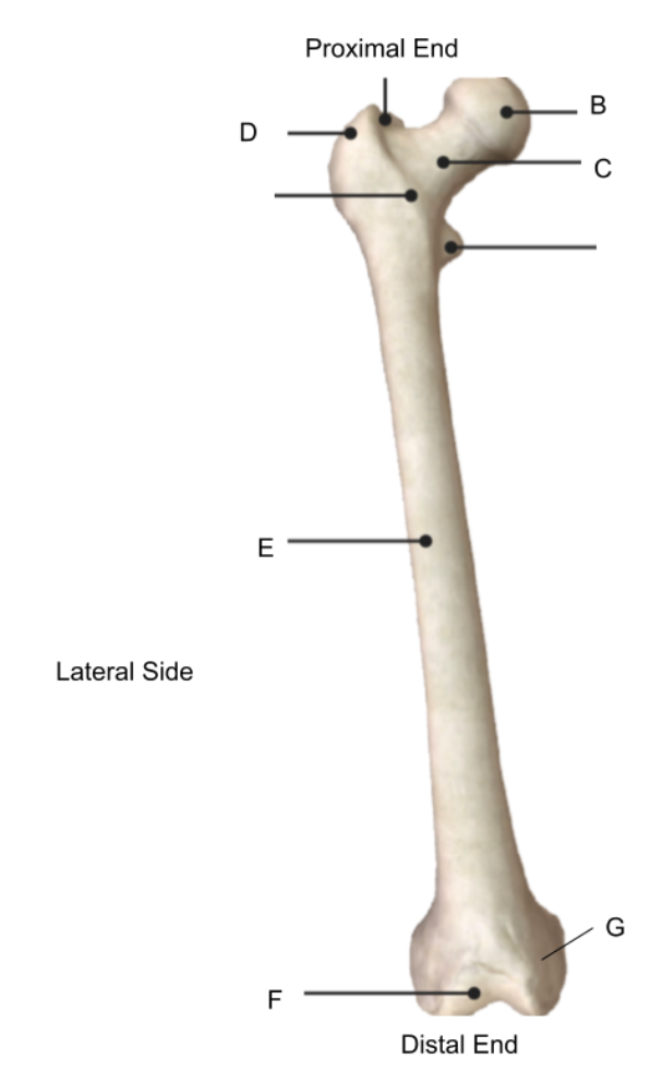
Match the structure with the correct letter
| Draggable item | arrow_right_alt | Corresponding Item |
|---|---|---|
trochanteric region | arrow_right_alt | B |
Intra-articular | arrow_right_alt | C |
femoral head | arrow_right_alt | E |
shaft | arrow_right_alt | G |
femoral neck | arrow_right_alt | D |
condyles | arrow_right_alt | F |
If bone tissue is built too quickly, it can become weak and easily fractured. This is due to an increase in
In certain metabolic diseases such as osteoporosis, increased
Put the stages of bone remodeling in the correct order
The fibrocartilage callus is gradually replaced by one made of spongy bone. This new mass is referred to as the bony callus. Osteoclasts and osteoblasts move to the area and multiply.
Over the weeks and months to come, the callus is remodeled with the help of osteoclasts and osteoblasts. The shape of the bones will gradually return to normal, and there will eventually be little evidence of the fracture.
Blood vessels that are ruptured during the break swell to form a mass called a hematoma. This mass forms between the broken bones. This clotting reduces the blood supply to many of the cells in the area of injury, and as a result, these cells die.
New capillaries begin to form into the clotted blood in the damaged area. Connective tissues cells form a mass of repair tissue called a fibrocartilage callus. This callus contains some cartilage, some bone, and collagen fibers. The combined mass closes the gap between the broken bones.
Match the description to the type of fracture repair
lower rate of infection
higher rate of infection
can only be used on a few types of fractures
can be used on all types of fractures
can only be used on the shaft or midel
requires a small incision
can be used on the proximal or distal end of a bone
requires a large incision
Plating
Nailing
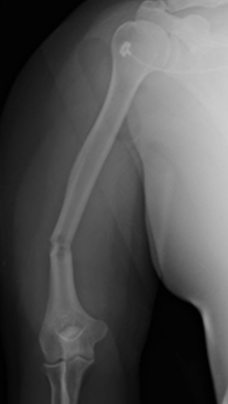
Nailing would be the best choice of repair due to only needing small incisions and the fracture being located on the shaft of the bone.
create an image of internal body structures such as tendons, muscles, joints, blood vessels, and internal organs; one of the few diagnostic imaging techniques that capture motion.
provide a two-dimensional image of the interior of the body; often used to provide images of the chest or broken bones
do not use radiation and can provide detail of very fine soft tissue; create images of the body using a large magnet and radio waves


X-Ray
Magnetic Resonance Imaging (MRI)
Ultrasound
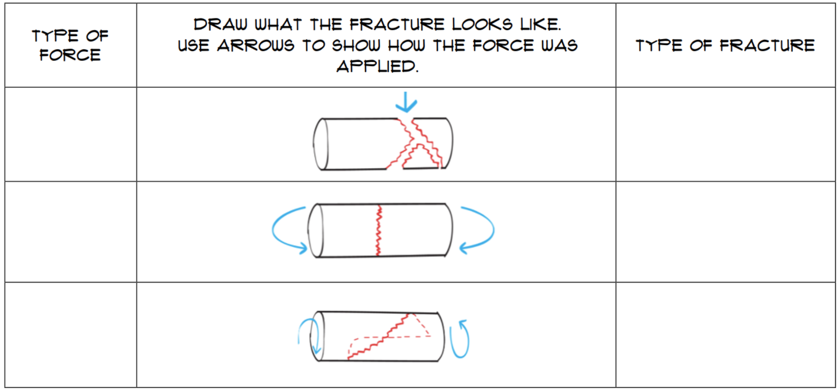
Type of force that causes this type of fracture
Type of force that causes this type of fracture
Type of force that causes this type of fracture
Type of fracture shown here
Type of fracture shown here
Type of fracture shown here