Drugi mogući odgovor:
proximal
superior
medial
lateral
superficial
inferior
anterior
posterior
deep
distal
patella
sternum
phalanges
fibula
skull
scapula
axial skeleton
appendicular skeleton
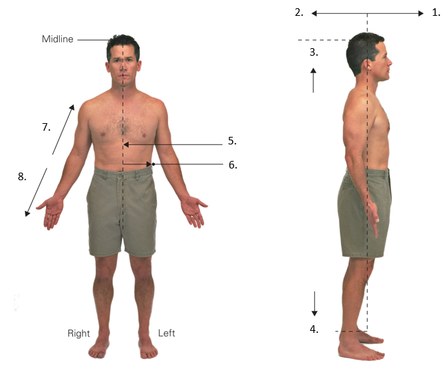
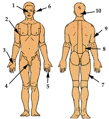
locate the anatomical regions
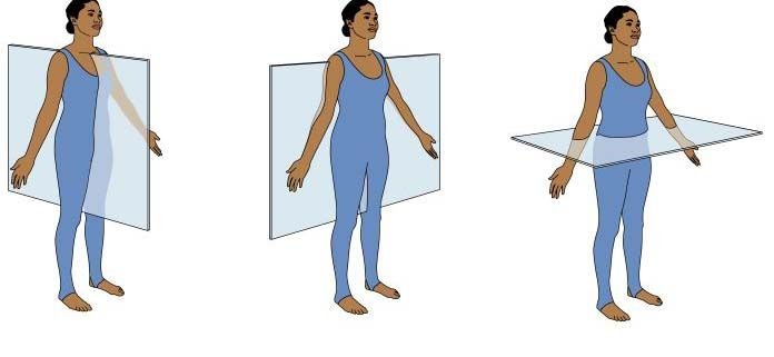
Click on the picture that shows a cross section through the coronal/frontal plane
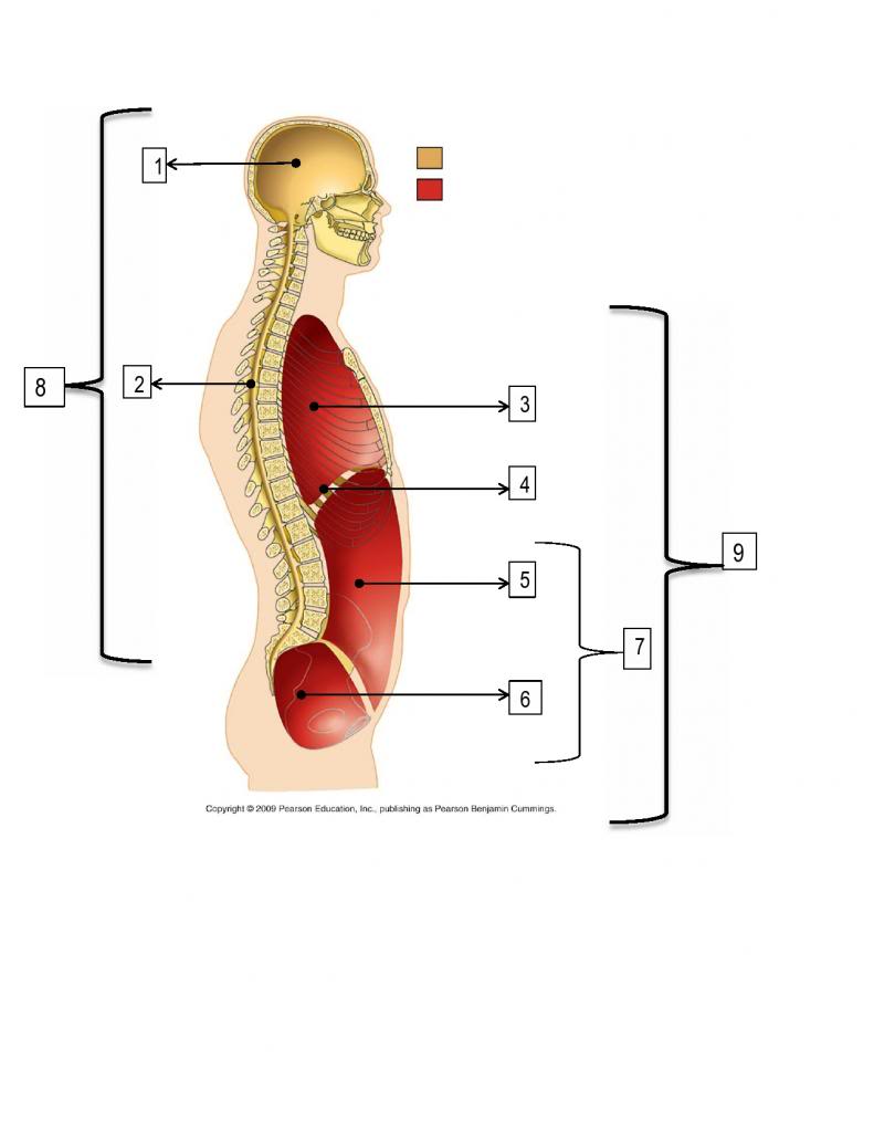
label the body cavities
put the hierarchy of tissues in order from smallest on top to largest on bottom
cell
organ system
organ
organisms
tissue
choose all that are the major tissue types
there are four different tissue types: bone,
organize the characteristics into either spongy or compact bone characteristics
haversian canal
bone marrow
trabecula
outer layer
inner layer
spongy
compact
| Stavka koja se može prevući | arrow_right_alt | Odgovarajuća stavka |
|---|---|---|
Short Bone | arrow_right_alt | made up of a layer of spongy bone between two thin layers of compact bone |
Flat Bone | arrow_right_alt | a bone that has a shaft and 2 ends and is longer than it is wide |
Irregular Bone | arrow_right_alt | bones are shaped roughly as a cube and contain mostly spongy bone |
Long Bone | arrow_right_alt | vary in shape and structure and therefore do not fit into any other category |
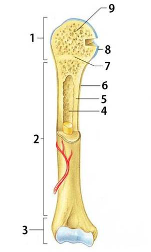
identify the different parts of a bone
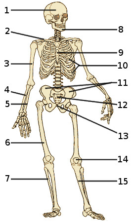
Identify the parts of the skeleton
classify the definitions and images
a fracture line that wraps around your bone and looks like a corkscrew
a bone that is broken in at least two places


occur when your bone is broken perpendicular to its length
a separation of two bones where they meet at a joint


dislocation
comminuted
transverse
spiral
put the steps of fracture repair in order from start (top) to finish (bottom)
Blood vessels that are ruptured during the break swell to form a mass called a hematoma.
fibrocartilage callus is gradually replaced by one made of spongy bone.
Over the weeks and months to come, the callus is remodeled with the help of osteoclasts and osteoblasts
New capillaries begin to form into the clotted blood in the damaged area. Connective tissues cells form a mass of repair tissue
