Note: Copy and Pasted answers will not be accepted.
Note: Copy and Pasted answers will not be accepted.
INTRODUCTION: In this lab, we will examine the anatomy of a rat. By dissecting the rat you will become familiar with basic anatomical directional terms, organs involved with each body system and the body cavities they are found in. The internal anatomy of a rat is very similar in construction to human anatomy with slight differences that will be explained in the rat dissection report. Please remember that your rat was once a living organism and should be treated with respect and care. Mutilation of your cadaver will result in a zero for the grade, removal from the lab area and the possibility of not partaking in another dissection during our course.
PURPOSE: To investigate the internal anatomy of a rat to determine basic anatomical directions, location of main organs and assignment of those organs to the organ systems. This lab will also introduce dissection techniques for those who have never performed a dissection.
MATERIAL:
Rat Cadaver - Rat Dissection Manual/Diagrams
Dissecting tray - Dissecting tools (scissors and blunt probe)
Camera (webcam or traditional)
SAFETY: You must wear goggles, apron, and gloves during the dissection. Always cut away from yourself or others. If you come into contact with any fluid from the rat, wash immediately with soap and water. If any other concerns arise during the dissection, flag down your instructor for assistance.
PRELAB: Posted on separately Google Classroom
Who were your group members? Include their name and role.
Dissector -
Photographer -
Lab Instructor -
Materials Management -
A.) Opening the Rat
Obtain dissecting tools, a rat, as well as a dissecting pan.
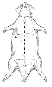
Use scissors to make the incisions in the rat. Hold the scissors so that they are horizontal with the rat’s abdomen. You may need to drain the fluids from the abdominal cavity. After puncturing the abdomen, hold the rat over the sink and allow it to drain.
After draining the rat, continue to cut making sure to not cut too deeply and keep the point of the scissors facing upward. Once the body cavity is opened, pin down the flaps to the dissecting tray or you may cut them off.
What two MAJOR body systems are predominantly involved during the opening the rat?
B.) Thoracic Cavity:
Locate the diaphragm which is a thin piece of muscle that separates the thoracic cavity from the abdominal cavity. Most inexperienced dissectors cut the diaphragm when opening the body cavities. If you cannot find the diaphragm after looking, flag down your teacher.
Locate the thymus gland positioned directly above the heart. This gland is triangular in shape and most inexperienced dissectors accidently tear it off mistaking it for muscle tissue. The thymus gland is involved in the development of T cells in the immune system.
Locate the heart. This organ will be found in the center of the thoracic cavity. Once the heart has been photographed,remove it from the cavity to see structures covered by its placement. *Note the four chambers: 2 atria & 2 ventricles.
Locate the trachea, bronchi, and lungs.
The trachea is a hard, ridged structure descending from the pharynx. You will need to cut high up on the throat and carefully move muscle to find it. The trachea will branch off in two tubes called bronchi, that lead to the large soft tissue of the left and right lungs.
Locate the bronchi by following the trachea down to where you removed the heart. The trachea will branch off in two tubes, the bronchi, that lead to the large soft tissue of the left and right lungs.
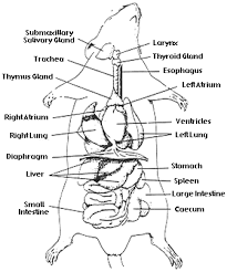
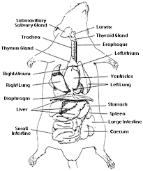
Label the following photo of a rat once you have completed this section of the lab. Label the following organs in the photo: diaphragm, thymus gland, heart, trachea, bronchi, and lungs. *****Please note that the numbers in the photo do not necessarily correspond with the labels you are being asked to add.
C.) Abdominal Cavity
Locate the diaphragm, again.
Locate the liver. The liver is the large dark purple/brown structure just underneath the diaphragm used for producing bile as well as storing glycogen and detoxifying the blood. You will not see a gallbladder in the rat!
Locate the esophagus. The esophagus moves down from the pharynx through the thoracic cavity and into the abdominal cavity ending at the stomach. It is next to the trachea and lacks the rings of cartilage found in the tracheaIf you cannot find it attached to the stomach you can also find it near, or behind the trachea. See image at right for reference.
Locate the stomach. The stomach is located underneath the diaphragm in the left side of the abdominal cavity. Note the layer of fat that hangs like an apron from the stomach. This is called the omentum.
Locate the spleen. This is a small dark purple/brown structure attached to the stomach. The spleen functions in the destruction of blood cells as well as blood storage.
Locate the pancreas. The pancreas is located in the tissue between the stomach and small intestine. It is brown and flat. Look for a thin, membranous (often bumpy) structure to find the pancreas. Don’t be afraid to move organs around to find the pancreas.
Locate the small intestine. The small intestine is thin and coiled as well as descends from the stomach. As you stretch out the small intestine you will see connective tissue holding the loops in place and serving as a pathway for blood vessels. This tissue is known as the mesentery.
Locate the large intestine/colon. This is the large tube extending from the small intestine to the anus.
Locate the cecum. This is the large sac most often confused with the large intestine. It is actually the point at which the small intestine becomes the large intestine.
Locate the kidneys by moving the small intestines, cecum and large intestines out of the way. There should be two on either side of the posterior wall of the abdominal cavity.
Locate the adrenal glands found on the superior region of the kidneys.
Locate the ureters descending down from each kidney.
Find the location where the ureters meet in the pelvic region. Dependent on the sex of the rat, you may need to move some structures aside. The urinary bladder will be a small, muscular sac in the most posterior section of the pelvic cavity.
What body systems did your group observe during the investigation through the abdominal cavity?
Use your laptop camera (or one of Mrs. Christman's cameras to take a photo of your rat's abdominopelvic cavity. Label the following organs in your photo: small intestine, large intestine, omentum, mesentery, spleen, kidney, and ureters.
Try not to cross your label lines because it is hard to tell what you are trying to point out. If a structure is not visible in your photo, please add a note.
D.) Pelvic Cavity
NOTE: You are dissecting only one rat; therefore, you will only see one sex during the actual dissection. You are, however, responsible for knowing the reproductive organs of both the male and female. Be sure to prepare your specimen so that it will be clear enough for another student to study. Also, make certain you examine a specimen of the sex that you did not dissect!
Male:
Locate the scrotum, a large sac of skin, muscle and connective tissue containing the testes on the exterior of the body just ventral to the anus. During non-breeding periods, the testes may be retracted into the abdominal cavity and the scrotum will not be enlarged. However, sperm cannot develop completely within the high temperatures of the abdominal cavity; the slightly lower temperature of the scrotum is necessary for the final stages of sperm formation. □
Carefully cut open the scrotum and locate the paired testes, which are oval. Around the outside of each testis is a C-shaped structure known as the epididymis, which is a very long, highly coiled tubule. □
Locate the urinary bladder if you haven’t already.
Female:
Locate the ovaries, which appear as a mass of follicles just posterior to the kidneys. They are often buried in a mass of fat, which should be carefully removed. The adult non-pregnant rat is usually in breeding condition with mature or well-developed ova in the ovaries.
Locate the right and left cornua (horns) of the uterus. The cornua unite to form the vagina.
Locate the urinary bladder if you haven’t already.
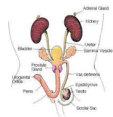
Examine this photo of the organs that are visible during your exploration of a MALE rat's pelvic cavity and external anatomy. Label the following organs: urinary bladder, epididymis, scrotum, and testis. If you do not have a male rat, take a look at a rat from another lab table. *****Please note that the numbers in the photo do not necessarily correspond with the labels you are being asked to add.
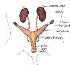
Take a photo of the organs that are visible during your exploration of a FEMALE rat's abdominopelvic cavity. Label the following organs in your photo: urinary bladder, ovary, uterine horns (cornua) and vagina. *****Please note that the numbers in the photo do not necessarily correspond with the labels you are being asked to add.
E.) Clean up
Obtain a plastic baggie. Put your name and period on it using a permanent marker.
Wrap your rat with paper towels sprayed with humectant and place in a plastic bag. Store per teacher instructions.
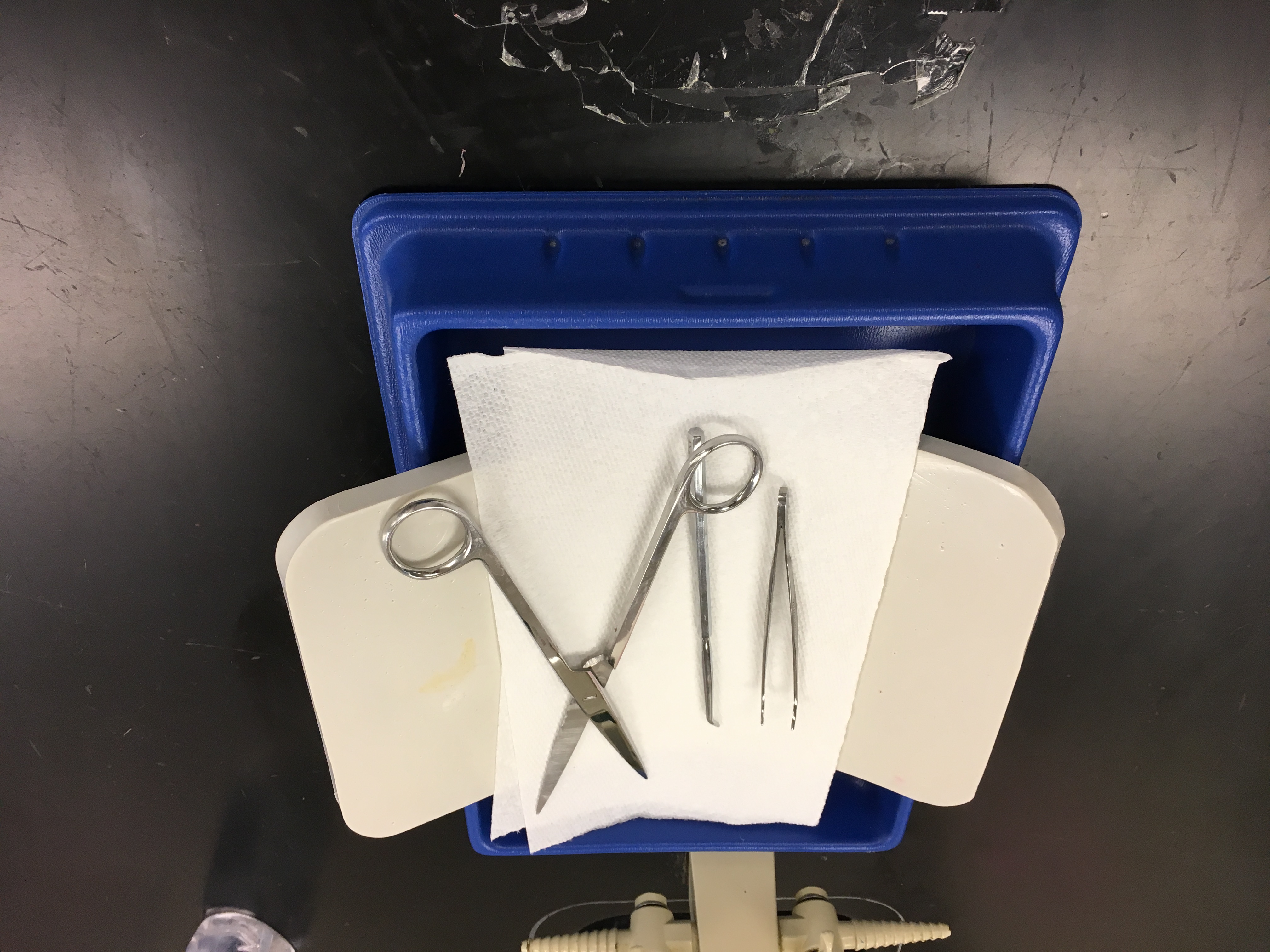
Rinse and disinfect trays and tools. Leave scissors to dry in the “open position” to prevent rusting.
Wipe down and disinfect the lab bench and clean out the sink.
*** This is what your tray should look like when you are finished.
Upload a photo of your CLEAN dissection tray and equipment next to the table number.
Which of the following the safety measures were required during the dissection?
Why was a rat a good choice for this dissection?
Identify the functions of the cardiovascular system.
Why does the rib cage have cartilage where the ribs and sternum attach? (You will have to research and cite your source.)
In your own words, define the term “lobe” when it is used to describe an anatomical feature.
The lungs have an unequal number of lobes on the left and right sides. Give a possible explanation which could account for this lack of symmetry based on the structure and function of the organs in this area.
How does the structure of the small intestine complement its function? (hint: sphincters/muscles, length, etc.)
Which structure serves as the anatomical divider between the thoracic and abdominal cavities? With which body system is it associated?
Match the organs with the processes they carry out.
large intestine
stomach
small intestine
cecum
digestion (breaks down food)
absorption
storage and fermentation of fiber-rich food
What body system(s) does the pancreas belong to?
What body system(s) does the spleen belong to?
Sequence the organs through which urine must travel in order to exit the body. Begin with the organ which filters the blood to form the urine.
kidney
urethra
ureter
bladder
The adrenal glands are not a part of the urinary system. What system do these belong to?
Human kidneys are said to be “retroperitoneal. ” What does this mean?
The renal arteries & veins are not considered part of the urinary system. Instead they are classified as part of the ______________________ system(s).
Which of the following organs are located in the thoracic cavity?
How does the STRUCTURAL difference between the human uterus and the rat uterine horn relate to the function of these organs?
What was your assigned role in the dissection for your lab group? What was the hardest part of the dissection from your point of view?