Ɛhia
1
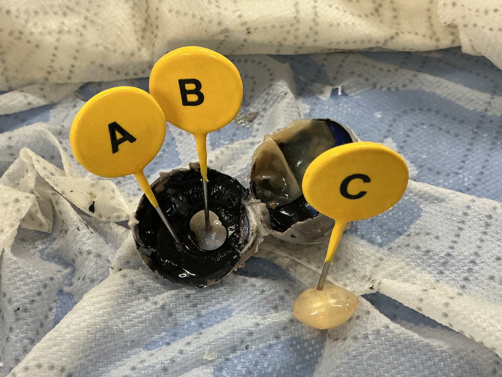
The structure labeled letter "A" helps send light into the eye. What is the name of this structure
The structure labeled letter "A" helps send light into the eye. What is the name of this structure
Ɛhia
1
Ɛhia
1
The structure labeled letter "A" helps send light into the eye. What is the name of this structure
What does the choroid layer provide? Check all that apply.
What is the primary function of rod cells in the eye?
What is the main function of the tapetum lucidum in a sheep eye?
What is the main function of the lens in a sheep eye?
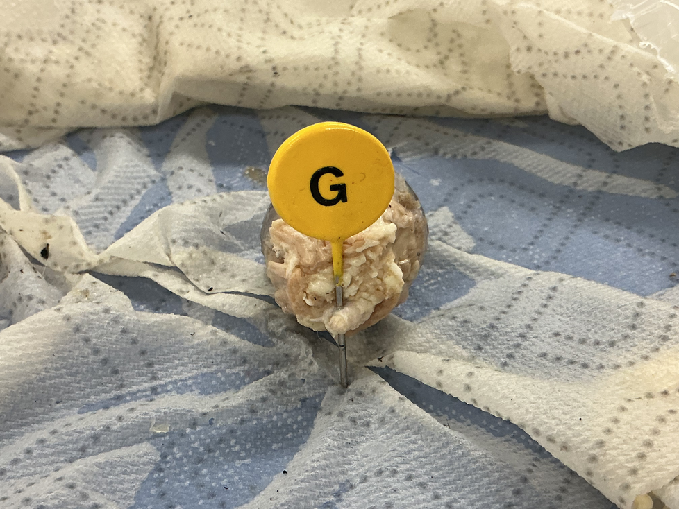
This is a view of the back side of the sheep eye. What is the structure labeled "G"?
Which type of photoreceptor is responsible for color vision?
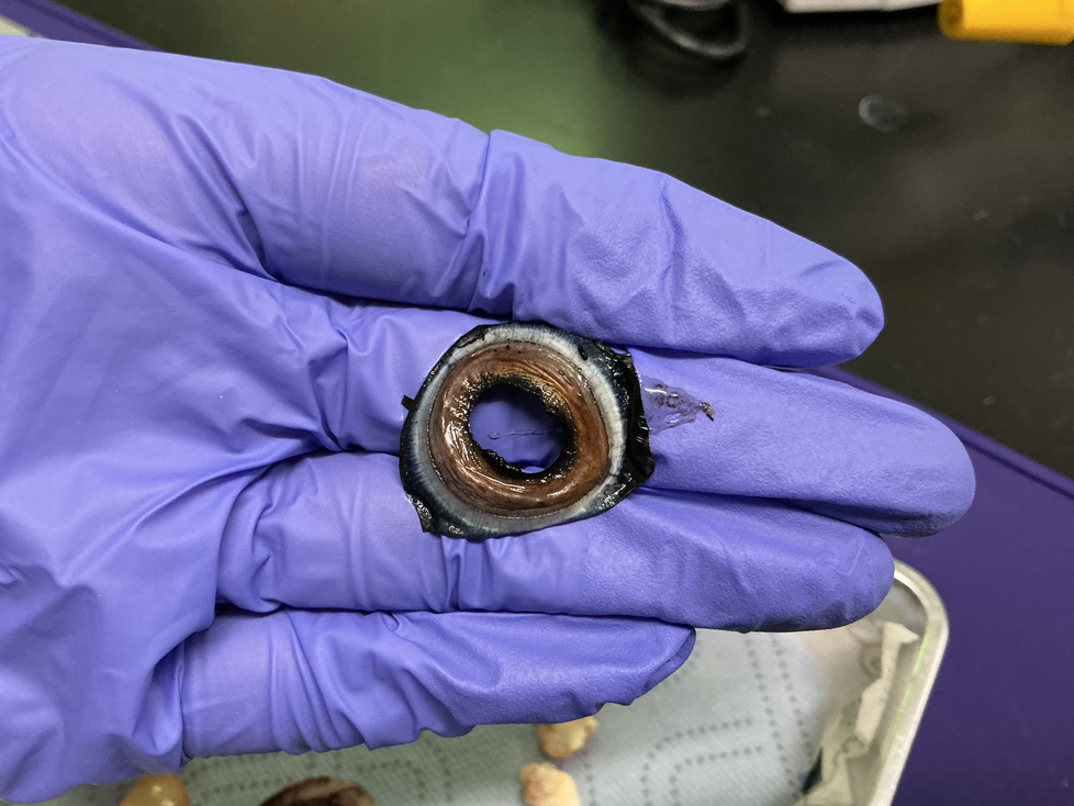
Is this a sclera?
What is the main function of the sclera?
These eye muscles allow the sheep to move its eyes from side to side. Check all that apply.
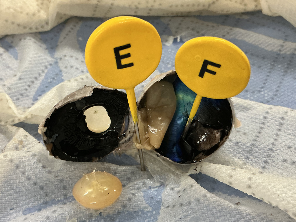
Match
| Draggable item | arrow_right_alt | Corresponding Item |
|---|---|---|
structure "F" | arrow_right_alt | found in both sheep and human eyes |
structure "E" | arrow_right_alt | found in sheep eyes, not in human eyes |
Where is the retina located?
What color is the iris of the sheep eye?
Answer(s) Confidence Tracker:

What is the structure labeled letter "B"?
The structure labeled letter "C" focuses images in the eye. You colored it light blue in your notebook model. What is the name of this structure?
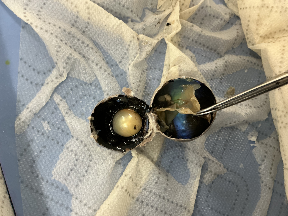
What is the name of the structure that the forceps is pulling on?
What is the function of the structure that the forceps is pulling on?
Where does the structure held by the forceps connect to the eye?
What is the name of the structure that looks like a pearl?
What is the function of the structure labeled "G"?