Copy of 09.15.20 (Sync and Async) - The Skull Bones (5/28/2026)
star
star
star
star
star
Last updated 3 months ago
20 Nsɛmmisa
1
1

1
1
1
1

1
1
1
1
1
1
1
1
1

1
1
1
1
1
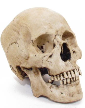
Objective: To identify the bones, processes, fossa, sutures, and other landmarks found in the human skull
Warm up #1
Articular cartilage is the cartilage present where bones articulate with other bones.
Brainstorm: what do you think the term 'articulate' means in anatomy?
Warm-Up #2
a. What do you know about the skull?
b. What do you want to know about it?
First, check out this simulation: https://www.visual-science.com/projects/skull/intaractive-app/
Which of the following perspectives of the skull are we able to see with this simulation?
For questions 4-6, you will be labeling a set of diagrams of the skull. Along with identifying the bones in the simulation above, you will be identifying various structures.
Use the text tool (not the scribble tool!) to write the names of the bones, then use the scribble tool to color/outline the bones as best as you can. Write the names of the bones in blue text.
Next, reference the textbook (not google!) to identify and label the structures provided with each question. Write the names of the structures in red text.

(there are times for it, but the time is not now!)
Use the simulation to identify, label (in blue text) and color the bones provided on the key above the diagram.
In addition to labeling and coloring the bones in the diagram, use the textbook to identify and label (in red text) the following structures:
squamous suture
coronal suture
lambdoid suture
external acoustic meatus
styloid process
mastoid process
mandibular condyle
(Notice that there are many more landmarks provided on the website. You are only responsible for knowing the locations of the landmarks listed above!)
Use the simulation above to identify, label (in blue text), and color the bones provided on the key above the diagram.
In addition to labeling and coloring the bones in the diagram, use the textbook to identify and label (in red text) the following structures:
supraorbital foramen
mental foramen
inforaorbital foramen
orbital surface of the sphenoid
greater wing of the sphenoid
(Notice that there are many more landmarks provided on the website. You are only responsible for knowing the locations of the landmarks listed above!)
Use the simulation above to identify, label (in blue text), and color the bones provided on the key above the diagram.
In addition to labeling and coloring the bones in the diagram, use the textbook to identify and label (in red text):
transverse palatine suture
mastoid process
foramen magnum
occipital condyle
zygomatic arch
anterior palatine foramen
median palatine suture
styloid process
(Notice that there are many more landmarks provided on the website. You are only responsible for knowing the locations of the landmarks listed above!)
Use the diagrams from questions 4-6 to answer questions 7-1516-20. Again - RESIST THE URGE TO GOOGLE!

(there are times for it, but the time is not now!)
Look back at your diagrams and identify the following general structures. Match the structure with its possible definition.
| Draggable item | arrow_right_alt | Corresponding Item |
|---|---|---|
process | arrow_right_alt | immobile joint between bones, usually allows for growth in childhood |
meatus | arrow_right_alt | a protuberance/ projection off of a bone, usually allowing for the attachment of muscle |
suture | arrow_right_alt | hole, usually allowing the passage of blood vessels, nerves, or ligaments |
foramen | arrow_right_alt | rounded surface of a bone, usually at an area of articulation |
condyle | arrow_right_alt | a tube-like passageway through a bone |
Match the following foramina (foramen) of the skull with the bone that they are located in.
| Draggable item | arrow_right_alt | Corresponding Item |
|---|---|---|
Infraorbital foramen | arrow_right_alt | Occipital bone |
Foramen magnum | arrow_right_alt | Frontal bone |
Mental foramen | arrow_right_alt | Maxilla |
Supraorbital foramen | arrow_right_alt | Mandible |
Which of the following bones contribute to the formation of the orbit of the eye?
There is one suture not pictured in the diagrams: The Sagittal suture.
Think back to your anatomical terminology lessons. Which of the following might best describe the location of the sagittal suture, and from which perspective do you think we would best be able to see it?
There are four prominent sutures of the adult skull. Identify the bones that join to form these sutures.
Frontal bone
Parietal bone(s)
Temporal bone(s)
Occipital bone(s)
Coronal suture
Lambdoid suture
Squamous suture
Sagittal suture
One of the bones you identified today, colored in yellow in the diagram below, is particularly hard to see from the lateral, inferior, and anterior perspectives. It is best seen from the superior perspective if a transverse cut is made through the skull:
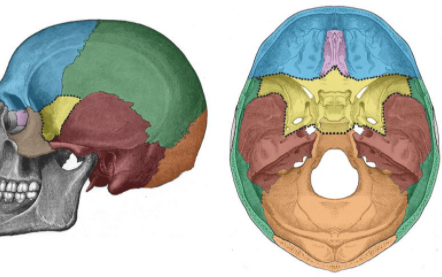
Which bone is this?
Six of the bones of the skull are unpaired. This means that there is only one of them.
Which of the following skull bones are unpaired?
Which bones of the skull would be considered parts of the cranium rather than the face?
Match the bones with their description
| Draggable item | arrow_right_alt | Corresponding Item |
|---|---|---|
Organs of hearing (internal ear) are located in and protected by these bones | arrow_right_alt | mandible |
This bone forms the lower jaw, including the chin | arrow_right_alt | zygomatic |
This bone forms the septum of the nose, diviging the nose into two nostrils | arrow_right_alt | nasal |
This bones form the roof of the mouth and contain sockets into which the upper teeth are set | arrow_right_alt | temporal |
These form the bridge of the nose | arrow_right_alt | parietal |
This name means "wall." The bones form most of the roof of the skull | arrow_right_alt | maxilla |
These are the cheek bones | arrow_right_alt | vomer |
Listen to the radiolab segment below. It takes about 20 minutes (yes, listen to the whole thing!). As you listen, look at the picture of the skull that the segment is about.
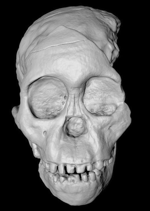
When you are done listening write a 4-5 sentence description of the Radiolab segment.
What is one interesting thing you learned during the radiolab segment?
What is one question you still have about the content in the radiolab segment?
The pictures below depict/display a condition called sagittal craniosynostosis
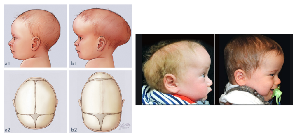
Using the medical terminology website:
break this term up into its components and
come up with a defintion for this condition.
(I WILL know if you use the website or not, and you won't get credit if you don't use it!)
Finally, watch the video below about Sagittal Craniosynostosis surgery.
a. Compare and contrast the two surgery options.
b. A child is diagnosed with sagittal craniosyntosis at two months old. Which procedure would you recommend and why?