Copy of 09.18.20 (Sync + Async) - The Appendicular Skeleton (5/28/2026)
star
star
star
star
star
Last updated 3 months ago
17 Nsɛmmisa
1
1

1
1
1
1
1
1
1

1
1
1
1
1
1
1
1
Objectives: Students will...
identify the bones, processes, fossa, sutures, and other landmarks found in the human arm, hand, and shoulder
identify the bones, processes, fossa, sutures, and other landmarks found in the human leg, foot, and hip
We will achieve these objectives while watching a series of videos, labeling and coloring a set of diagrams, and answering a set of questions
1-2
A Little Note from your Teacher:
Please, please, PLEASE - resist the urge to simply google the answers to these questions. I know that the videos contain a lot of information and may feel overwhelming. Just remember that learning about the structures of the body is what anatomy is all about! Also, remember that you are not required to memorize these structures - rather, we are practicing the skill of transferring information from a carefully selected media source (video) to diagrams and questions. I was very intentional with the sources I chose - please use them.

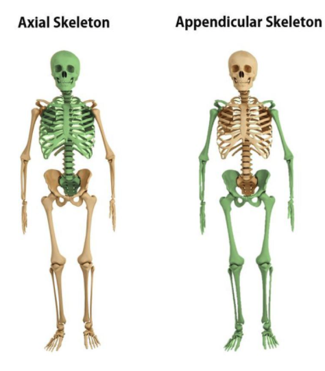
Using the first diagram above, compare and contrast the axial and appendicular skeleton.
Warm-Up: Match the bone structure with its description. You may use the textbook for help if you like!
| Draggable item | arrow_right_alt | Corresponding Item |
|---|---|---|
crest | arrow_right_alt | rounded surface of a bone |
head | arrow_right_alt | something that "sticks off" of the bone |
tubercle | arrow_right_alt | ridge |
foramen | arrow_right_alt | small, rounded process |
condyle | arrow_right_alt | elongated basin |
fossa | arrow_right_alt | prominent, rounded surface of a bone |
process | arrow_right_alt | hole through a bone |
You will be working in groups for questions 4-9
Quality Control will make sure all group members agree on answer before moving on to next question.
Manager shares their screen and makes sure group stays on same question and stays on task.
Spokesperson is contacts Ms. Whitney in the main Google meet if your group has a question.
Timekeeper makes sure group completes task and returns to whole class Google meet on time.
First, watch the video below. As you watch, color the bones in the diagram below (under the other videos):
Bones of the Arm:
Next, watch the three videos below. After each video, scroll to the bottom of this question and label the structures discussed during the video for that bone.
Humerus:
(before you go on - scroll down and label the diagram with the terms that apply to the humerus!)
Radius:
(before you go on - scroll down and label the diagram with the terms that apply to the radius!)
Ulna:
(before you go on - scroll down and label the diagram with the terms that apply to the ulna!)
acromion process
clavicle
coracoid process
capitulum
greater tubercle
lesser tubercle
head of humerus
coronoid fossa
trochlea
radial tuberosity
styloid process of radius
head of radius
head of ulna
styloid process of ulna
(the diagram is of the right arm facing anteriorly)
What are two interesting / fun facts you learned about the arm and shoulder bones?
Watch the video below:
Label the diagram below using the following terms:
scapular spine
vertebral margin
coracoid process
acromion process
glenoid fossa (on diagram twice!)
axillary margin
(the lateral aspect is a left scapula, and the posterior aspect is a right scapula)
Watch the video below about the structures that make up the elbow. You do NOT have to watch the whole thing, and please don't worry about the movements (we'll be talking about those in a few weeks!)
Label the diagram below using the following terms:
humerus
radius
ulna
olecranon process
olecranon fossa
Reference your diagrams to match the bones/features below with their description.
| Draggable item | arrow_right_alt | Corresponding Item |
|---|---|---|
part of the humerus that articulates with the scapula | arrow_right_alt | olecranon fossa |
long, oblique ridge on the posterior surface of the scapula that extends from the lateral border of the acromion | arrow_right_alt | olecranon process |
depression on the proximal end of the ulna that articulates with the trochlea of the humerus | arrow_right_alt | glenoid fossa |
condyle of the humerus that articulates with the ulna | arrow_right_alt | coronoid fossa |
A fossa on the distal, posterior surface of the humerus | arrow_right_alt | acromion process |
condyle of the humerus that articulates with the radius | arrow_right_alt | greater tubercle |
fossa on the distal, anterior surface of the humerus | arrow_right_alt | semi-lunar (trochlear) notch |
edge of the scapula nearest the armpit | arrow_right_alt | capitulum |
large process on the head of the humerus | arrow_right_alt | trochlea |
The proximal process of the ulna | arrow_right_alt | head of humerus |
depression on the scapula that articulates with the humerus | arrow_right_alt | scapular spine |
edge of the scapula nearest the vertebral column | arrow_right_alt | lesser tubercle |
process on the scapula that articulates with the clavicle | arrow_right_alt | vertebral margin |
small process just above the surgical neck of the humerus | arrow_right_alt | axillary margin |
Extension:
Have you ever hit your "funny bone?" Did you know that the funny bone isn't a bone at all?
Examine the following picture as you answer the questions below:
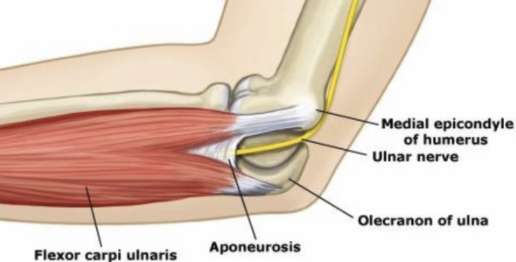
1. What do you think is actually "hit" when someone hits their "funny bone"
2. One can only hit their "funny bone" when their elbow is bent. Explain why this is.
3. What is a more scientific way to express "Owe! I hit my funny bone!"?
Watch the three videos below. After each video, scroll to the bottom of this question and color the bones AND label the structures discussed during the video for that bone.
Pelvis
(before you go on - scroll down and label the diagram with the terms that apply to the pelvis!)
Femur
(before you go on - scroll down and label the diagram with the terms that apply to the femur!)
Tibia
(before you go on - scroll down and label the diagram with the terms that apply to the tibia!)
Fibula
(before you go on - scroll down and label the diagram with the terms that apply to the fibula!)
inominate
sacrum
coccyx
head of femur
neck of femur
lateral condyle
medial condyle
lesser trochanter
intertrochanteric line
greater trochanter
cnemial crest
head of fibula
tarsals
metatarsals
phalanges
symphysis pubis
patella
medial malleolus
tibial tuberosity
What are two interesting / fun facts you learned about the leg and hip bones?
Watch the video below, then label the diagram using the following terms. Finally, color the inominate diagram with the provided colors.
acetabulum
sacro-iliac joint
symphysis pubis
obturator foramen
iliac crest
coccyx
Watch the video below, then color and label the diagram using the following terms.
calcaneus
talus
cuboid
navicular
intermediate cuneiform
lateral cuneiform
Reference your diagrams to match the bones/features below with their description.
| Draggable item | arrow_right_alt | Corresponding Item |
|---|---|---|
thin, lateral bone of the lower leg | arrow_right_alt | talus |
oblique ridge between the trochanters on the anterior surface of the femur | arrow_right_alt | cuboid |
the most superior bone of the inominate | arrow_right_alt | intertrochanteric line |
fossa on the inominate that articulates with the femur | arrow_right_alt | iliac crest |
large process lateral to the neck of the femur | arrow_right_alt | acetabulum |
large bump found on the medial aspect of the distal tibia | arrow_right_alt | inominate |
longest, strongest bone in the leg | arrow_right_alt | pubis |
most posterior bone of the inominate (your "sit bones") | arrow_right_alt | symphysis pubis |
the crest of bone on the anterior part of the tibia | arrow_right_alt | patella |
joint between the sacrum and ilium of the inominate | arrow_right_alt | ilium |
structure consisting of both inominates, sacrum, and coccyx | arrow_right_alt | fibula |
The tarsal bone that articulates with the most lateral metatarsal | arrow_right_alt | femur |
process just inferior to the neck of the femur on the medial surface | arrow_right_alt | sacro-iliac joint |
the largest tarsal bone | arrow_right_alt | lesser trochanter |
one half of the pelvic girdle | arrow_right_alt | head of femur |
the uppermost ridge of the inominate | arrow_right_alt | ischium |
unattached bone in the knee | arrow_right_alt | pelvic girdle |
part of the femur that fits into a depression on the inominate | arrow_right_alt | greater trochanter |
the tarsal bone that articulates with the tibia | arrow_right_alt | calcaneus |
joint between pubic bones on the anterior pelvis | arrow_right_alt | cnemial crest |
large bump found on the proximal, anterior aspect of the tibia | arrow_right_alt | medial malleolus |
the anterior bone of the obturator foramen | arrow_right_alt | tibial tuberosity |
The image below was obtained from a patient who was in a car accident. The patient was in the passenger seat with their feet up on the dashboard when they hit another car head on.
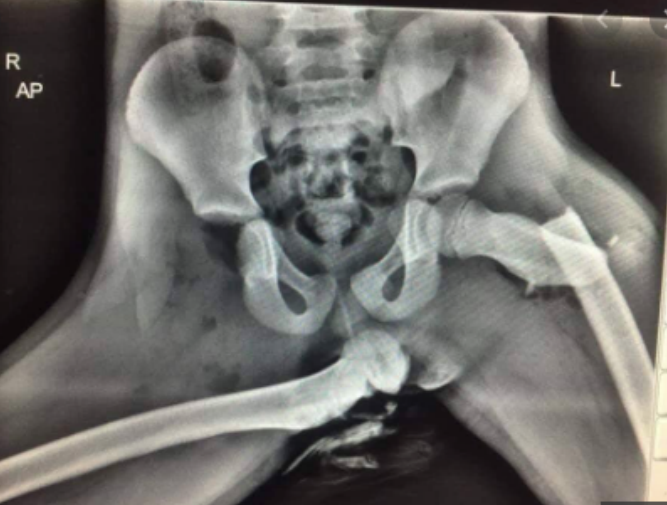
Use this picture to answer questions
What is the best description of what happened to this patient?
Do some quick research on what types of surgeries might help this patient. Attach a diagram of one (or more) of the procedures that you might recommend for them if you were an orthopedic surgeon.
Go to this link: Whack a Bone (you might need to change your computer's settings to allow flash to run)
Pick at least one of the three games on the website. Play the game, then upload a screenshot of your results in the space below. (if you don't know how to do this, look it up!)