Copy of 10.02.20 (Sync + Async) - Sliding Filament Model (5/28/2026)
star
star
star
star
star
Last updated 3 months ago
22 Nsɛmmisa
1
1
1
1
1
1
1
1
1
1
1
1
1
1
1
1
1
1
1
1
1
7
Objectives: Students will...
differentiate between the proteins, enzymes, and other biochemicals important in the Sliding Filament Model of muscle contraction
differentiate between the steps involved in the Sliding Filament Model of muscle contraction
write a love letter or story about various proteins involved in muscle contraction
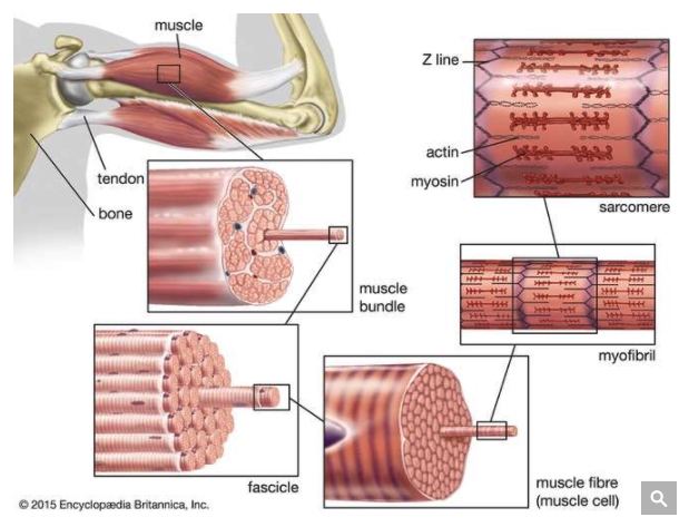
Put the following muscle structures in order from largest to smallest.
Fascicle
Muscle fiber (muscle cell)
Muscle
Myofibril
Categorize the following substances as proteins or ions.
Tropomyosin
Ca2+
Na+
Actin
Myosin
Titin
Troponin
Protein
Ion
First, identify the following as true or false:
Myofibrils are smaller than sarcomeres.
Then, draw a picture justifying your answer.
The picture below displays a simplified version of a single sarcomere.
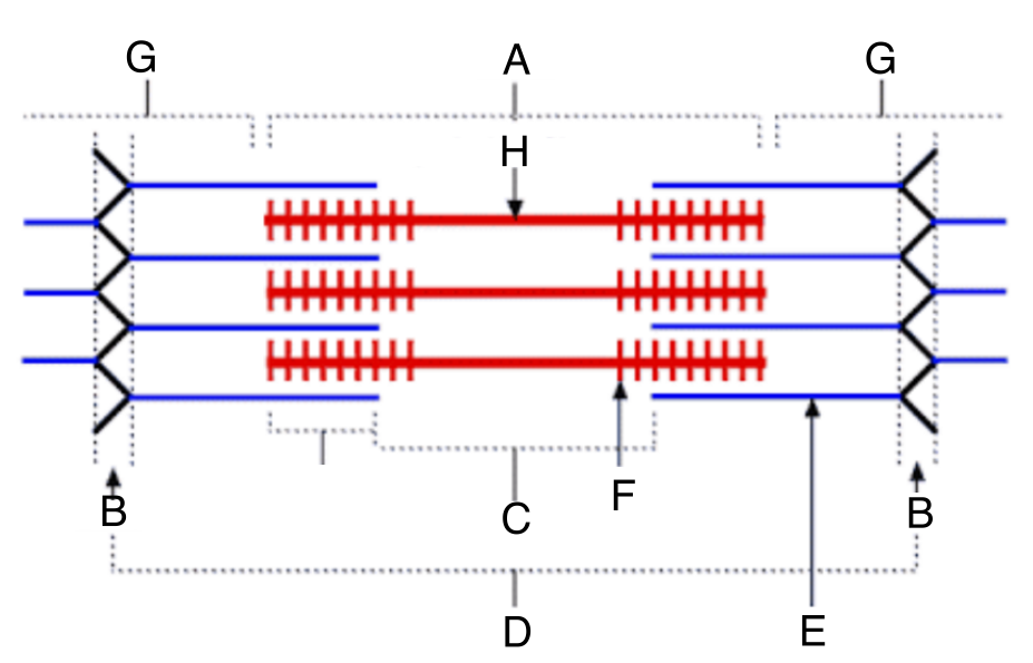
Match the terms with the correct letter on the diagram.
| Draggable item | arrow_right_alt | Corresponding Item |
|---|---|---|
G | arrow_right_alt | A band |
C | arrow_right_alt | Z line |
A | arrow_right_alt | H zone |
H | arrow_right_alt | Sarcomere |
D | arrow_right_alt | Thin filaments |
B | arrow_right_alt | Thick filaments |
E | arrow_right_alt | I band |
F | arrow_right_alt | Myosin heads |
Which of the following best describes the difference between the A band and the I band?
Now let's talk about thick and thin filament in a little more detail. Examine the picture of thick and thin filaments below while answering questions 6-9.
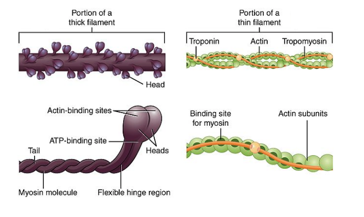
Match the following proteins with the structure they are on
Myosin
Actin
Tropomyosin
Troponin
Thick filaments
Thin filaments
Which of the following best describes thick filaments?
What binds at each of these sites on myosin?
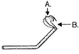
| Draggable item | arrow_right_alt | Corresponding Item |
|---|---|---|
B | arrow_right_alt | Actin |
A | arrow_right_alt | ATP |
Compare and contrast troponin and tropomyosin.
What questions do you still have about Part 1: Skeletal Muscle Structure?
Skeletal muscle only contracts when it is activated by the nervous system. Nervous system cells, like muscle cells, are excitable - this means that they can send messages within their cells using electrical signal. If not for the electrical signals of your nervous and muscle cells, we would not exist as we do today.
Communication between the nervous cells and muscle cells requires another form of communication: chemical communication. Neurotransmitters are the chemicals used to relay messages from a nervous cell to a muscle cell. While we will learn more about neurotransmitters when we get to the nervous system next semester, a basic understanding of this signaling is important in understanding how muscles contract.
Watch the following video about the neuromuscular junction. Remember that we have not yet talked about the nervous system and that we will learn more about neuron structure and action potentials next semester. For now, focus on the terms provided below.
| Draggable item | arrow_right_alt | Corresponding Item |
|---|---|---|
Acetylcholine (ACh) | arrow_right_alt | Space between neuron and muscle cell |
ACh Receptors | arrow_right_alt | Neurotransmitter released from neuron |
Ca2+ | arrow_right_alt | Proteins on the membrane of the muscle cell that bind to ACh |
Synaptic cleft | arrow_right_alt | Ions that enter the muscle cell when enough ACh has attached to the muscle cell receptros |
Na+ | arrow_right_alt | Ions that enter the neuron and trigger the release of ACh |
Compare and contrast the roles of Na+ and Ca2+ in muscle contraction.
What questions do you still have about Part 2: The Neuromuscular Junction?
Watch the following video on muscle contraction.
(Note: You may watch the whole thing, but the beginning is review. He'll start talking about skeletal muscle contraction at 1:45)
As you watch, match the terms below with the best description.
| Draggable item | arrow_right_alt | Corresponding Item |
|---|---|---|
Tropomyosin | arrow_right_alt | Protein |
Titin | arrow_right_alt | Long, "strink-like" protein blocking the binding sites on actin |
Troponin | arrow_right_alt | Binds to the protein troponin in order to expose myosin binding site on actin |
Myosin | arrow_right_alt | Small protein attached to tropomyosin when muscle is at rest; binds to Ca2+ during muscle contraction |
Ca2+ | arrow_right_alt | Protein holding the thick filaments in place |
Describe the relationship between tropomyosin, troponin, myosin, and actin
Ok - Let's talk more about ATP. Examine the picture below, focusing on Adenosine Triphosphate (ATP), Adenosine Diphosphate (ADP), and Phosphate (P).
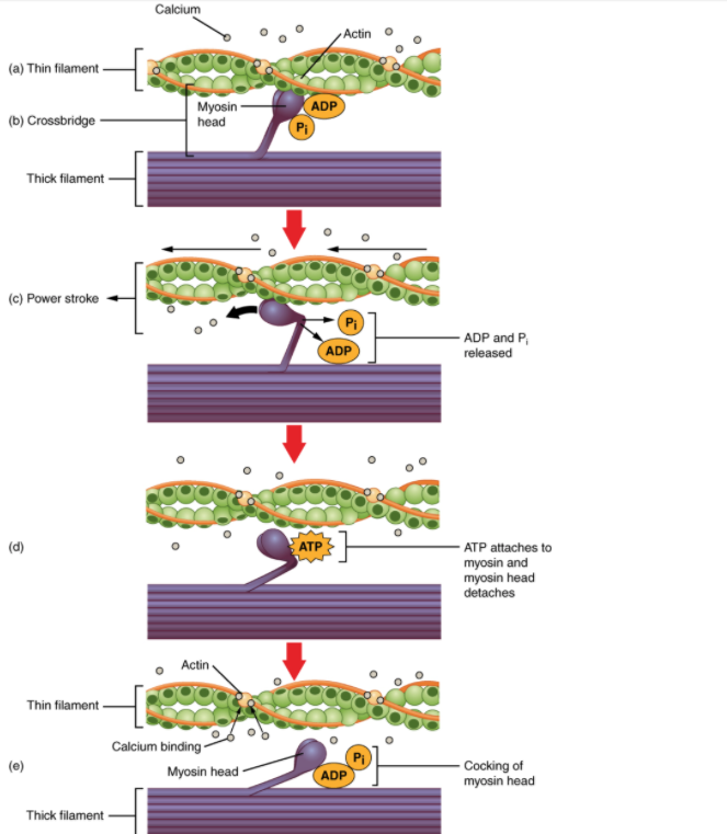
Which of the following statements about the role of ATP, P, and ADP in muscle contraction are accurate?
Think back to this simplified description of ATP hydrolysis that we saw the other day (left) and compare it to the more complex, accurate description on the right.
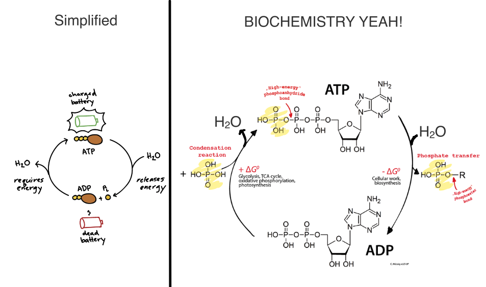
Which of the following best describes ATP hydrolysis?
What questions do you still have about Part 3: The Art of Muscle Contraction
WITHOUT LOOKING AT YOUR NOTES: Put the following steps of muscle contraction in the correct order. The order should go from before a contraction --> contraction
ATP reacts with water, forming ADP and phosphate. The energy created is used to move the myosin head back to its original position.
Myosin heads rotate towards the center of the sarcomere, bringing the Z-lines closer together
Calcium ons (Ca2+) bind to troponin molecules
Sodium ions (Na+) enter the plasma membrane of the muscle cell. Potassium ions (K+) exit the plasma membrane of the muscle cell
The shape of troponin is altered, causing tropomyosin to move and expose the actin binding sites on the thin filaments
Acetylcholine crosses the synaptic cleft and binds to receptors on the muscle fiber
The myosin heads of the thick filaments attach to the actin binding sites on the thin filaments, forming a cross bridge
ADP and phosphate are released fom their binding sites on the myosin heads
Calcium ions (Ca2+) are released from the sarcoplasmic reticulum of the muscle cell
ATP binds to myosin, disconnecting the crossbridge
What is still confusing about the Sliding Filament Model?
Refer back to Section 10.2 or Section 10.3 of your textbook and fill in your confusion.
How did the textbook help? What are you still confused about?
Before you move on, go back to #19 and try to re-sequence the steps. You can use your notes this time!
Then, re-watch the Crash Course:
Do you need to resequence again? What things make sense now that didn't make sense before?
Role: Some type of protein involved in muscle contraction (Acton, Myosin, Tropomyosin, Troponin)
Format: Love Letter
Topic: Your feelings towards another protein (or proteins) involved in muscle contraction. You should discuss / refer to the following in your letter:
actin
myosin
tropomyosin
troponin
ATP
ADP and P
water
Ca2+
thick filaments
thin filaments
sarcomeres
myofibril
Z-lines
mitochondria