2
a.) According to the diagram there are
b.) The nerve,
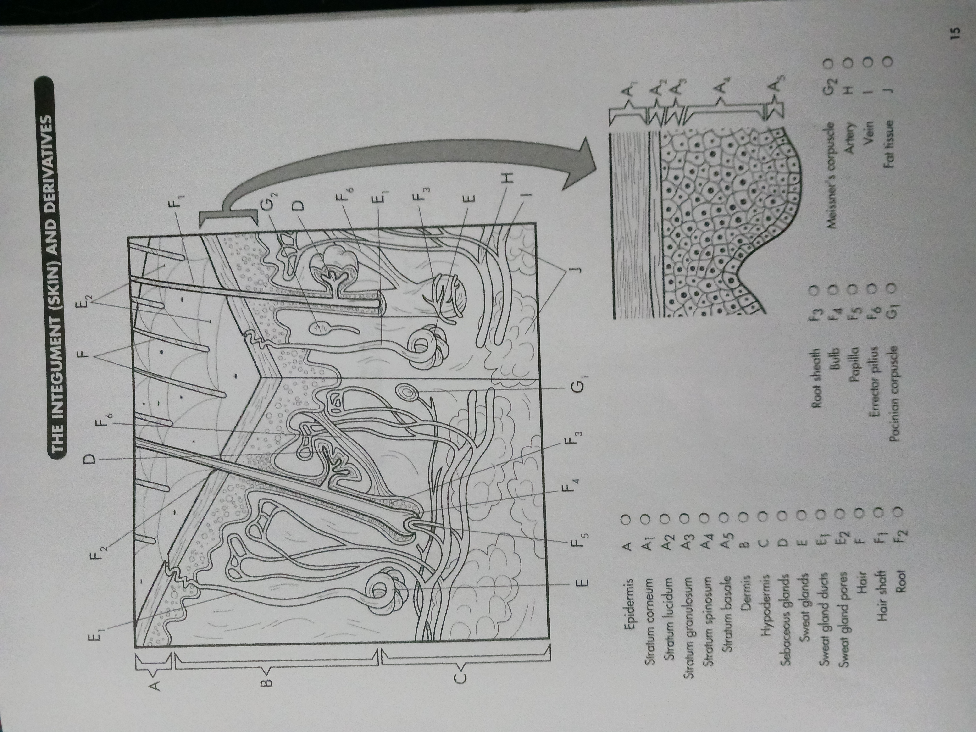
The integumentary system is composed of the integument (skin) and its derivatives, including the hairs, sweat glands, and oil glands. As the largest organ of the body, the skin provides protection to the body and performs other functions.
On a structural basis, the skin is composed of three parts. At its surface the epidermis (A), which is outlined by a bracket in the diagram. The next deep layer contains connective tissue and is called the dermis (B). Then comes a subcutaneous layer called the hypodermis (C).
Returning to the epidermis, we note the detailed view and identify five layers of tissue. Moving from the surface downward, the most superficial (close to the surface) layer is the stratum corneum (A1). This is a layer of flat, dead cells filled with the protein keratin. The layer protects against heat, pathogenic microorganisms, chemicals, and light. Next comes the stratum lucidum (A2). Clear, flat cells with a prekeratin substance called eleidin are found here. The layer is found primarily in the palms of the hands and soles of the feet.
The next lower layer of the epidermis is the stratum granulosum (A3), which contains flattened cells containing the substance keratohyalin. Later, this material will become keratin. The next deeper layer is a very large layer called the stratum spinosum (A4). Keratin is produced in many of these cells.
The deepest layer is the stratum basale (A5). It is a single layer of cuboidal and columnar cells that undergo mitosis and become the cells of the more superficial (those above) layers. The layer is also called the stratum germinativum.
a.) The skin provides
b.) The skin is comprised of three primary layers, from the deepest to the most surface layer, they are
c.) The dermis contains
a.) The
b.) The answer to the first blank in 2a is made up of two types of cells, they are
c.) Moving downward from the surface, the
d.) Located primarily on the palms of the hands and soles of the feet, the
e.) The stratum spinosum is the layer of the epidermis where
f.) Because it is the lowest layer of the epidermis the
g.) The outermost layer, the
The dermis contains many fibers of collagen together with various kinds of cells. The most superficial region of dermis is the papillary region with fingerlike projections in the epidermis. The remainder of the dermis is the dermal layer.
Within the dermal layer are a number of sebaceous glands (D). These oil glands are generally connected to hair follicles, as indicated on the diagram. Their secretion is an oily substance called sebum. Other glands in the dermis are the sweat glands (E), also called sudoriferous glands. These glands deliver their watery secretions (sweat) to sweat gland ducts (E1), which lead to sweat gland pores (E2). Sweat performs excretory functions by delivering metabolic waste products to the skin surface for removal. It also helps regulate body temperature.
Hairs (F) are epidermal growths distributed in varying amounts and textures throughout the body. The superficial part of the hair is the shaft (F1), projecting above the body surface. The portion penetrating into the dermis is the root (F2). The root of the hair is covered by the root sheath (F3), which is a continuation of the epidermis, as the diagram indicates. At the base of the hair follicle is the enlarged hair bulb (F4). And indentation called the papilla (F5) contains connective tissues and blood vessels to provide nourishment to the hair.
At the side of the hair follicle is a specialized smooth muscle called the errector pilius (F6). This muscle contracts during stress and pulls the hair to the upright position. It can be seen in both hair fibers on the diagram.
Many types of nerve receptors are located within the dermis. While there are many types of receptors in the body, only two receptors will be discussed here. First is the Pacinian corpuscle (G1). This nerve receptor detects vibrations and heavy touch sensations and sends impulses off to the brain. Meissner's corpuscles (G2) detect light touch sensations and dispatch impulses for interpretation.
a.) Numerous types of cells are found with fibers of
b.) Featuring fingerlike projections that extend into the epidermis, the
c.) Generally connected to hair follicles,
d.) Releasing watery secretions through ducts that lead to skin pores, seat glands are also known as
e.) The visible part of hair above the skin is known as the
f.) The
g.) The nerve that senses light touch and distributes signals for interpretation is the
In the hypodermis, the diagram displays a number of nerves, as well as the blood supply of the integumentary system. An artery (H) carries blood to the skin, while a vein (I) carries blood away. Much fat tissue (J) is found in the hypodermis to provide cushioning to the skin. The underlying muscles are below the hypodermis.
a.) Found in the hypodermis, a large amount of
b.) In the integumentary system, a
a.) According to the diagram there are
b.) The nerve,