Log in
Sign up for FREE
arrow_back
Library

Copy of Cardiac Conduction System Reading (5/28/2026)

star
star
star
star
star
Last updated about 3 hours ago
5 questions
Note from the author:
1
1
Use the reading from each section to help you with these questions.

The questions will not immediately tell you if you are correct or not.
  • Once you submit your first attempt, check your score.
  • If below an 80%, reattempt them.
  • All correct answers are saved from the first attempt.
  • Up to 4 total attempts - 85% is highest score possible on attempts 2-4.
Use the reading from each section to help you with these questions.

The questions will not immediately tell you if you are correct or not.
  • Once you submit your first attempt, check your score.
  • If below an 80%, reattempt them.
  • All correct answers are saved from the first attempt.
  • Up to 4 total attempts - 85% is highest score possible on attempts 2-4.
12
Question 1
1.

Atrioventricular (AV) Node

The atrioventricular (AV) node is a second clump of specialized myocardial conductive cells, located in the inferior portion of the right atrium within the atrioventricular septum. The septum prevents the impulse from spreading directly to the ventricles without passing through the AV node. There is a critical pause before the AV node depolarizes and transmits the impulse to the atrioventricular bundle. This delay in transmission is partially attributable to the small diameter of the cells of the node, which slow the impulse. Also, conduction between nodal cells is less efficient than between conducting cells. These factors mean that it takes the impulse approximately 100 ms to pass through the node. This pause is critical to heart function, as it allows the atrial cardiomyocytes to complete their contraction that pumps blood into the ventricles before the impulse is transmitted to the cells of the ventricle itself. With extreme stimulation by the SA node, the AV node can transmit impulses maximally at 220 per minute. This establishes the typical maximum heart rate in a healthy young individual. Damaged hearts or those stimulated by drugs can contract at higher rates, but at these rates, the heart can no longer effectively pump blood.
9

Atrioventricular Bundle (Bundle of His), Bundle Branches, and Purkinje Fibers

Arising from the AV node, the atrioventricular bundle, or bundle of His, proceeds through the interventricular septum before dividing into two atrioventricular bundle branches, commonly called the left and right bundle branches. The left bundle branch has two fascicles. The left bundle branch supplies the left ventricle, and the right bundle branch the right ventricle. Since the left ventricle is much larger than the right, the left bundle branch is also considerably larger than the right. Portions of the right bundle branch are found in the moderator band and supply the right papillary muscles. Because of this connection, each papillary muscle receives the impulse at approximately the same time, so they begin to contract simultaneously just prior to the remainder of the myocardial contractile cells of the ventricles. This is believed to allow tension to develop on the chordae tendineae prior to right ventricular contraction. There is no corresponding moderator band on the left. Both bundle branches descend and reach the apex of the heart where they connect with the Purkinje fibers. This passage takes approximately 25 ms.

The Purkinje fibers are additional myocardial conductive fibers that spread the impulse to the myocardial contractile cells in the ventricles. They extend throughout the myocardium from the apex of the heart toward the atrioventricular septum and the base of the heart. The Purkinje fibers have a fast inherent conduction rate, and the electrical impulse reaches all of the ventricular muscle cells in about 75 ms. Since the electrical stimulus begins at the apex, the contraction also begins at the apex and travels toward the base of the heart, similar to squeezing a tube of toothpaste from the bottom. This allows the blood to be pumped out of the ventricles and into the aorta and pulmonary trunk. The total time elapsed from the initiation of the impulse in the SA node until depolarization of the ventricles is approximately 225 ms.
9
Question 4
4.

Question 5
5.

Sinoatrial (SA) Node

Normal cardiac rhythm is established by the sinoatrial (SA) node, a specialized clump of myocardial conducting cells located in the superior and posterior walls of the right atrium in close proximity to the orifice of the superior vena cava. The SA node has the highest inherent rate of depolarization and is known as the pacemaker of the heart. It initiates the sinus rhythm, or normal electrical pattern followed by contraction of the heart.

This impulse spreads from its initiation in the SA node throughout the atria through specialized internodal pathways, to the atrial myocardial contractile cells and the atrioventricular node. The internodal pathways consist of three bands (anterior, middle, and posterior) that lead directly from the SA node to the next node in the conduction system, the atrioventricular node (see Figure 19.18). The impulse takes approximately 50 ms (milliseconds) to travel between these two nodes. The relative importance of this pathway has been debated since the impulse would reach the atrioventricular node simply following the cell-by-cell pathway through the contractile cells of the myocardium in the atria. In addition, there is a specialized pathway called Bachmann’s bundle or the interatrial band that conducts the impulse directly from the right atrium to the left atrium. Regardless of the pathway, as the impulse reaches the atrioventricular septum, the connective tissue of the cardiac skeleton prevents the impulse from spreading into the myocardial cells in the ventricles except at the atrioventricular node. Figure 19.19 illustrates the initiation of the impulse in the SA node that then spreads the impulse throughout the atria to the atrioventricular node.
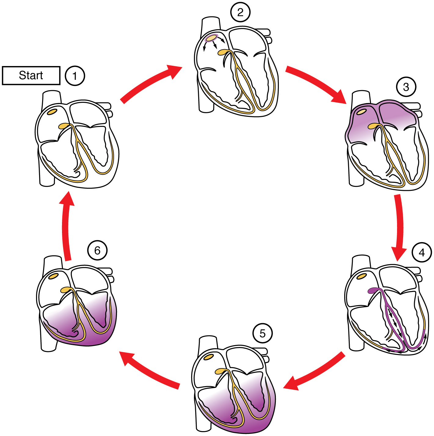

Figure 19.19 Cardiac Conduction (1) The sinoatrial (SA) node and the remainder of the conduction system are at rest. (2) The SA node initiates the action potential, which sweeps across the atria. (3) After reaching the atrioventricular node, there is a delay of approximately 100 ms that allows the atria to complete pumping blood before the impulse is transmitted to the atrioventricular bundle. (4) Following the delay, the impulse travels through the atrioventricular bundle and bundle branches to the Purkinje fibers, and also reaches the right papillary muscle via the moderator band. (5) The impulse spreads to the contractile fibers of the ventricle. (6) Ventricular contraction begins.

The electrical event, the wave of depolarization, is the trigger for muscular contraction. The wave of depolarization begins in the right atrium, and the impulse spreads across the superior portions of both atria and then down through the contractile cells. The contractile cells then begin contraction from the superior to the inferior portions of the atria, efficiently pumping blood into the ventricles.
a.) The sinoatrial (SA) node is located in the superior and posterior walls of the _______ .

b.) The SA node is known as the _______ of the heart due to its highest rate of _______ .

c.) The impulse from the SA node spreads throughout the atria through specialized _______ .

d.) The internodal pathways consist of _______ bands.

e.) It takes approximately _______ milliseconds for the impulse to travel from the SA node to the _______ .

f.) _______ conducts the electric impulse from the right to left atrium.

g.) The delay at the atrioventricular node allows the atria to _______ before the impulse is transmitted to the _______ bundle.

h.) The wave of depolarization begins in the right atrium and spreads across the _______ portions of both atria.

i.) The electrical event of depolarization triggers _______.
Question 2
2.
a.) The atrioventricular (AV) node is located in the _______ portion of the _______ .

b.) The _______ prevents the impulse from spreading directly to the _______ without passing through the AV node.

c.) There is a _______ before the AV node depolarizes and transmits the impulse.

d.) It takes approximately _______ milliseconds for the impulse to pass through the AV node.

e.) The pause allows the atrial _______ to complete their _______ before the impulse is transmitted to the ventricles.

f.) The AV node can transmit impulses maximally at _______ per minute with extreme stimulation by the SA node.
Question 3
3.
a.) The atrioventricular bundle, also known as the _______ arises from the AV node.

b.) The answer to 'a' proceeds through the _______ before dividing into _______ .

c.) The _______ supplies the left ventricle, while the _______ supplies the right ventricle.

d.) Portions of the right bundle branch are found in the _______ and supply the right papillary muscles.

e.) The _______ fibers spread the impulse to the _______ cells in the ventricles.

f.) The contraction of the ventricles begins at the _______ and travels toward the _______ of the heart.
Which structures are involved in the AV septum?
Atrioventricular Node
Purkinje Fibers
Bundle of His
Sinoatrial Node
Which structures are responsible for electrical impulses in the ventricles?
Purkinje Fibers
Bundle Branches
Bundle of His
Sinoatrial Node