Copy of Mitosis Cell Identification - Virtual Lab (5/28/2026)
star
star
star
star
star
Last updated 3 months ago
13 Nsɛmmisa
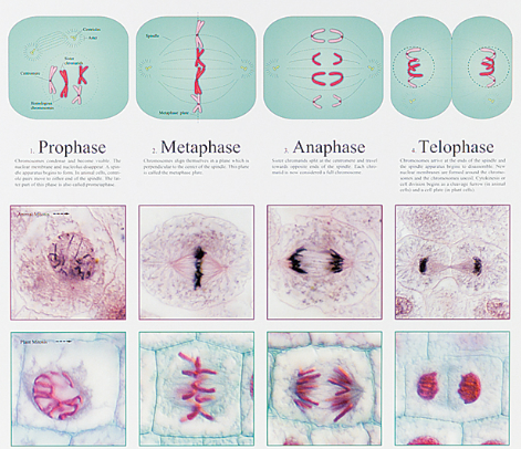
Part 1: Notes - Class Work
10
Part 2: Identification - Individual Work
Cytokinesis is the process that follows mitosis (or meiosis) in cell division, during which the cytoplasm of a parent cell is divided into two daughter cells; in animal cells, the cell membrane pinches in; in plant cells, a cell plate forms.
Chromatin is a complex of DNA and proteins found in the nuclei of eukaryotic cells; usually not visible; DNA is loosely wrapped around the proteins.
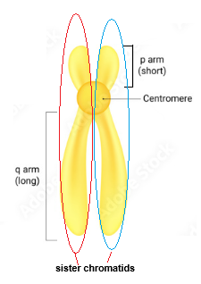
Chromosome is a long, thread-like structure made of DNA and proteins that carries genetic information; essentially, it is condensed chromatin (DNA is tightly wrapped around proteins, taking up less space in the nucleus); usually visible.
Centromere - the region of a chromosome where the two sister chromatids are joined together. It plays a crucial role during cell division, as it is the point where spindle fibers attach to pull the sister chromatids apart.
Sister chromatids are two identical copies of a single chromosome that are connected at the centromere. They are formed during DNA replication and are separated during cell division, ensuring that each daughter cell receives an identical set of chromosomes.
P-arm is the shorter arm of a chromosome, located above the centromere. The designation "p" stands for "petit," which means small in French.
Q-arm is the longer arm of a chromosome, located below the centromere. The designation "q" comes from the next letter in the alphabet after "p."
Submit the Mitosis Cell Identification Notes you created in the this section.
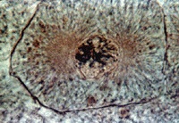
What stage of mitosis is this cell in?
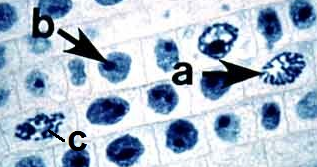
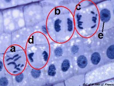
What stage of mitosis are cell "a" and "c" in?
Cell "a":
Cell "c":
Cell "b" has a fully intact nucleus that includes a nucleolus. Is this cell in any stage of mitosis?
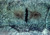
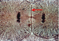
What stage of mitosis is this cell in?
This is an animal cell; what structure is being used to divide the daughter cells?
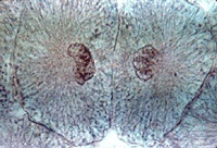
What stage of mitosis is this cell in?
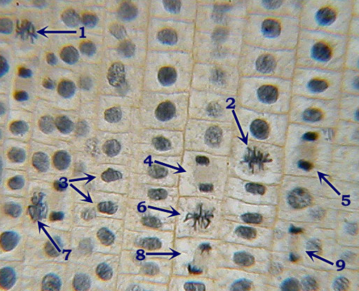
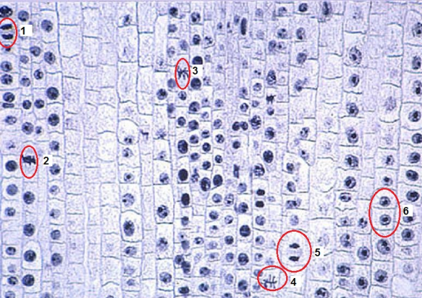
Identify each of the following labeled cells:
Cell 1
Cell 2
Cell 3
Cell 4
Cell 5
Cell 6
Cell 8
Identify each of the following labeled cells:
Cell 1
Cell 2
Cell 3
Cell 4
Cell 5
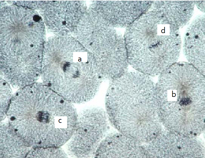
Identify each of the following labeled cells:
Cell a
Cell b
Cell c
Cell d
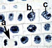
Identify each of the following labeled cells:
Cell a
Cell b
Cell c
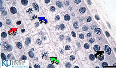
Identify each of the following labeled cells:
Red arrow
Blue arrow
Green arrow
Black arrow
Identify each of the following labeled cells:
Cell a
Cell b
Cell c
Cell d
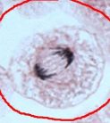
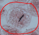
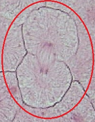
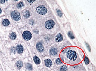
What stage of mitosis is the cell in the red circle?
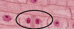
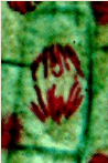
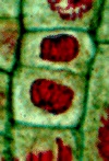
What stage of mitosis is this cell in?
What structure is separating the daughter cells that are forming?
What process is separating the daughter cells?