Log in
Sign up for FREE
arrow_back
Library

Copy of Reading Guide: Cell Division in Eukaryotes (5/28/2026)

star
star
star
star
star
Last updated about 3 hours ago
1 question
Note from the author:
10

Cell Division in Eukaryotes

Though cell division in all cells results in one cell becoming two cells, cell division in eukaryotic organisms is very different from that in prokaryotes, mainly because of the many chromosomes in the nuclei of eukaryotic cells. Cell division in eukaryotic organisms is necessary for development, growth, and repair of the organism. Just as in binary fission, eukaryotic cell division ensures that each resulting daughter cell receives a complete copy of the organism’s entire genome. Remember that all of an organism’s DNA must be present in each somatic, or body, cell. This DNA contains the information necessary for that cell to perform its functions, and to give that organism its traits. Therefore, prior to cell division, the eukaryotic cell’s complete genome must be copied, a process known as DNA replication, ensuring that each daughter cell receives a complete set of the genome. Prior to cell division, the cell's organelles are also duplicated. Now the cell is ready to divide. Cell division occurs at the end of an eukaryotic cell's cell cycle.

Eukaryotic cell division occurs in two major steps:
  1. The first step is mitosis, a multi-phase process in which the nucleus of the cell divides. During mitosis, the nuclear membrane breaks down and later reforms. The chromosomes are also sorted and separated to ensure that each daughter cell receives a diploid number of chromosomes. In humans, that number of chromosomes is 46 (23 pairs). Mitosis is described in greater detail in Cell Cycle: Mitosis (Advanced). Because the DNA has replicated prior to mitosis, the two nuclei that result from mitosis are genetically identical.
  2. The second major step is cytokinesis. As in prokaryotic cells, the cytoplasm must divide. Cytokinesis is the division of the cytoplasm in eukaryotic cells, resulting in two genetically identical daughter cells.

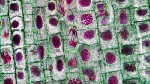
Figure 2: Shown are cells in various stages of their cell cycle. Numerous dividing cells are evident.

The formation of gametes, an organism’s reproductive cells, such as sperm and egg cells, involves a completely different method of cell division, called meiosis. This cell division ensures that each gamete receives a haploid number (half the amount) of chromosomes.
Question 1
1.

Attach your completed copy of the Reading Guide, here.

Exit Ticket

  • Before attempting this, review the reading guide.
  • 15 multiple choice questions
  • 7 minutes
  • Do not navigate off of the exit ticket window; otherwise, it will lock.
  • If you score below a 70% on the first attempt, you may do it once more (max of 70% on attempt two).
https://assessment.peardeck.com/author/tests/verid/680bb30603c2dae529266e7f

DNA, Chromosomes, and Genes

DNA contains the information necessary to make proteins, direct a cell’s activities, and give an organism its traits. Obviously, it is a very important molecule. Actually , in human cells, DNA is organized into 46 molecules called chromosomes.

The information in DNA is organized into structural units scattered along the length of the DNA molecule. These units are known as genes. A gene contains the information necessary to encode an RNA molecule or a protein. A diploid human cell has about 44,000 genes; two copies of each of about 22,000 genes. So, a single DNA molecule contains hundreds to thousands of genes. Through the process of gene expression, which involves transcription and translation, different cell types use the information in different genes to make different proteins. This process gives different cell types distinct activities. Thus, a liver cell will have many different proteins than a kidney cell, giving the two cells types distinct activities. When a cell is using the information within a gene, the segment of DNA containing that gene is unwound as chromatin, exposing the double helix to the cell machinery needed to use that information.

Prior to cell division, the DNA must duplicate itself in a process called DNA replication. This ensures that each resulting cell receives a complete set of the organism’s genome. But how is the replicated DNA divided up evenly? This will be discussed in an additional concept.

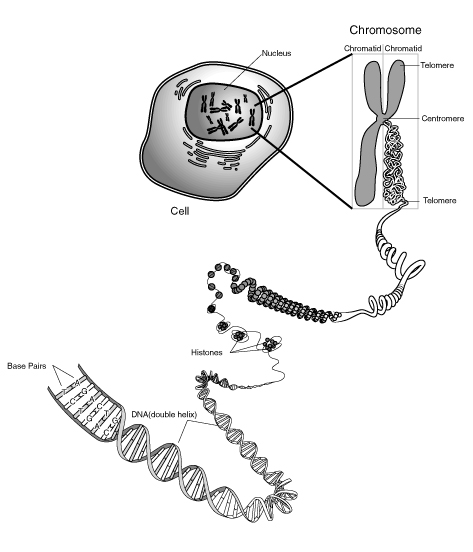
What guarantees that each new cell will receive a complete set of DNA? It was the identification of chromosomes that allowed this process to be characterized. As a eukaryotic cell prepares to divide, the DNA and associated proteins (histones) coil into a chromosome (Figure below). The DNA copies itself prior to this process, so the chromosome that forms consists of two identical chromatids, known as sister chromatids, identical copies of DNA. The two chromatids are attached at a region called the centromere. The chromatids separate from each other when the nucleus divides just prior to cell division. Thus, each new cell that results after cell division will have the complete complement of genetic material, identical to the original, or parent, cell. In human cells, this amounts to 46 chromosomes. These chromosomes come in pairs (one from each pair inherited from each parent). So these 46 chromosomes are actually two sets of 23 chromosomes each.


Figure 2: The DNA double helix wraps around proteins and tightly coils a number of times to form a chromosome. This figure shows the complexity of the coiling process. The chromosome comprises two identical, or sister, chromatids held together by a centromere.

Each human somatic cell (a body cell, or every cell other than a gamete) normally has two sets of chromosomes, one set inherited from each parent. These cells are said to have a diploid number of chromosomes. Each set contains 23 chromosomes, for a total of 46 chromosomes. Each chromosome differs in size, from about 250 million nucleotide pairs on the largest chromosome (chromosome #1) to less than 50 million nucleotide pairs on chromosome #22. Each chromosome contains a specific set of genes, as well as regulatory elements and other nucleotide sequences, making each chromosome essential to survival.

Homologous Chromosomes

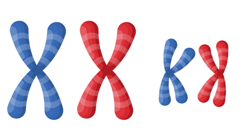
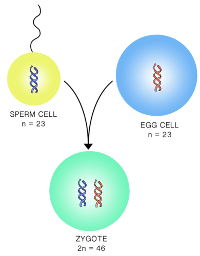
Each pair of chromosomes consists of two chromosomes that are similar in size and shape. They contain the same genes at the same loci, though they may have different alleles. These pairs of chromosomes are known as homologous chromosomes or homologues. Upon fertilization, a zygote is formed (Figure below). A zygote is the first cell of a new individual. In humans, a zygote contains 23 pairs (or two sets) of chromosomes. Any cell containing two sets of chromosomes is said to be diploid. The zygote forms from the fusion of two haploid gametes. A haploid cell contains one set of chromosomes. In humans, a haploid gamete contains 23 chromosomes. Biologists use the symbol n to represent one set of chromosomes, and 2n to represent two sets.

Figure 3: Homologous chromosomes form a pair, one from each parent. Homologous chromosomes are similar in size and shape, and contain the same genes, though they may have different alleles. Alleles are alternative forms of the same gene. This diagram represents two pairs of homologous chromosomes.


Figure 4: Upon fertilization a diploid zygote is formed. In humans, a zygote has 46 chromosomes, 23 inherited from each parent. The gametes, sperm and eggs, are haploid cells, with 23 chromosomes each.

Sex Chromosomes

In humans, each set of chromosomes contains 22 autosomes and 1 sex chromosome. Autosomes are chromosomes that are not directly involved in determining the sex of an individual. The sex chromosomes contain genes that determine the sex of an individual.

Whereas autosomes are found as homologous pairs in somatic cells, sex chromosomes come in two different sizes, shapes, and contain different genes. In many organisms, including humans, the sex chromosomes are known as the X and Y chromosomes. The Y chromosome contains genes that cause male development. Therefore, any individual with a Y chromosome is male, and a male will have both an X and Y chromosome (XY). Females, without a Y chromosome, will have two X chromosomes (XX). As females have two X chromosomes, they must pass an X chromosome to all of their children. As males have both an X chromosome (inherited from their mother) and a Y chromosome, they can give either chromosome to their children. If a child inherits a Y from his father, he will be male; if a child inherits an X from her father, she will be female. It therefore is the male gamete that determines the sex of the offspring.

Mitosis

Mitosis is the division of the cell's nucleus, the final step before two daughter cells are produced. Mitosis begins immediately at the conclusion of interphase, specifically at the end of the G2 phase. The cell enters mitosis as it approaches its size limitations. Four distinct phases of mitosis have been recognized: prophase, metaphase, anaphase, and telophase, with each phase merging into the next one (Figure below).
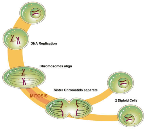
Figure 2: Mitosis is the phase of the eukaryotic cell cycle that occurs between DNA replication and the formation of two daughter cells. What happens during mitosis? During mitosis, the nucleus divides, paving the way for two cells to be produced after cell division, each with a complete makeup of genetic material.

The Phases

Prophase

Prophase is the first and longest phase of mitosis, see Figure below. During prophase, the chromatin (DNA) coils up into visible chromosomes, each made up of two sister chromatids held together by the centromere. Also during this phase, the nucleolus disappears, and the spindle begins to form from the centrioles. Most eukaryotic cells contain structures known as centrosomes, consisting of a pair of centrioles. During prophase, the centrioles begin to move to opposite ends, or poles, of the cell. As the centrioles migrate, the fiber-like spindle begins to elongate between the centrioles. The spindle is a thin, cage-like structure made out of microtubules. In plant cells, the spindle forms without centrioles. The spindle plays an essential role moving chromosomes and in the separation of sister chromatids.
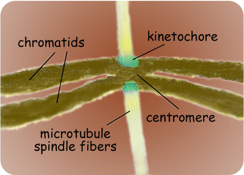
Figure 3: The spindle starts to form during prophase of mitosis. Kinetochores on the spindle attach to the centromeres of sister chromatids.

Metaphase

During metaphase, the centromeres of the chromosomes line up along the metaphase plate or equatorial plane, in essence the approximate middle of the cell. This orientation of the chromosomes at the equator of the cell helps to ensure proper chromosome separation. This alignment allows the spindle fibers to correctly pull the chromatids to either pole of the cell, resulting in separation of sister chromatids from a chromosome, see Figure below.
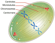

Figure 4: Chromosomes, consisting of sister chromatids, line up at the equator (metaphase plate) of the cell during metaphase.

Anaphase

Anaphase is the phase in which the sister chromatids separate. The sister chromatids are pulled apart by the shortening of the microtubules of the spindles, similar to the reeling in of a fish by the shortening of the fishing line. One sister chromatid moves to one pole of the cell, and the other sister chromatid moves to the opposite pole. This process occurs when the proteins that bind sister chromatids together are cleaved, resulting in unattached identical chromosomes, essentially separate daughter chromosomes. These separate chromosomes are pulled apart by shortening spindle fibers, and pulled toward the centrosomes to which they are attached. At the end of anaphase the spindle fibers degrade. At this time, each pole of the cell has a complete set of chromosomes, identical to the amount of DNA at the beginning of G1 of the cell cycle.


Telophase

Telophase is essentially the opposite of prophase and prometaphase. The chromosomes begin to unwind back into chromatin in preparation to direct the cell's metabolic activities. A new nucleus forms around each set of chromosomes. This is followed by cytokinesis, the division of the cytoplasm, resulting in two genetically identical cells, ready to enter G1 of the next cell cycle. The phases of mitosis are summarized in Figure below.
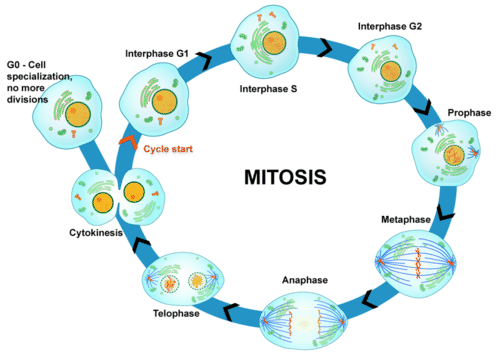
Figure 5: Mitosis in the Eukaryotic Cell Cycle. Mitosis is the multi-phase process in which the nucleus of a eukaryotic cell divides. In this diagram, prometaphase is not included as a separate phase, but incorporated into prophase.
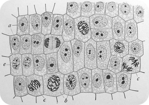

Figure 6: This is a representation of dividing plant cells. Cell division in plant cells differs slightly from animal cells as a cell wall must form. Note that most of the cells are in interphase. Can you find examples of the different stages of mitosis?

Cytokinesis

Cytokinesis is the final step in cell division. It often occurs concurrently with telophase, though it is a separate process. Cytokinesis (Figure below) differs between plant and animal cells. In animal cells, the plasma membrane pinches inward along the cell's equator until two cells are formed. Specifically, a cleavage furrow containing a contractile ring develops in approximately the middle of the cell (similar to the position of the metaphase plate), essentially pinching off the two nuclei and forming separate cells. In plant cells, a cell plate forms along the cells equator. A new membrane grows along each side of the cell plate, with a new cell wall forming on the outside of each new membrane.

At the end of cytokinesis, each daughter cell has a complete copy of the genome of its parent cell. The end of cytokinesis marks the end of the M-phase, the end of one cell cycle, and the beginning of G1 and interphase of the next cell cycle.
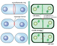
Figure 7: Cytokinesis is the final stage of eukaryotic cell division. It occurs differently in animal (left) and plant (right) cells.