Copy of 09.09.20 (Sync) - Bone Functions, Classifications, and Structure (5/28/2026)
star
star
star
star
star
Last updated 3 months ago
21 Nsɛmmisa
1
1

1
1

1
1
1
1
1
1

1
1
1
1
1
1
1
1
1
1
1
In this lesson, you will:
justify the importance of bone tissue in the overall workings of the human body
identify and distinguish between the gross structures of bones
identify and distinguish between the cells located within compact and spongy bone
Warm-Up #1:
What do you already know about the skeletal system?
Warm-Up #2:
What do you want to know about the skeletal system?
On your own: Brainstorm
What are the functions of the skeletal system? Think about the system as a whole rather than individual bones.
According to the video, which of the following is NOT a function of the skeletal system?
Welcome to your new group! We will be staying in these groups for a few weeks.
Think about your answers to the following questions, then share with your group members:
1. Your name
2. Your favorite season (and why)
3. Your favorite type of food (i.e. Italian, Sushi, Barbecue, etc.)
4. One thing you hope to learn about the skeletal system over the next three weeks.
In the space below: who are your group members?
Draw a picture of something that you have in common with one or more of your group members.
Discuss with your group: Who will take each role for today?
Reader will read all informational text and questions out loud to the group.
Manager makes sure group stays on same question and finishes on time.
Spokesperson is ready to share group's answers with the whole class.
Timekeeper makes sure group completes task and returns to whole class Google meet on time.
What is your role today?
The 206 bones of the adult human body can all be classified as one of the following: Long, Short, Flat, Irregular, or Sesamoid.
With your group, take a minute to look at the following table:
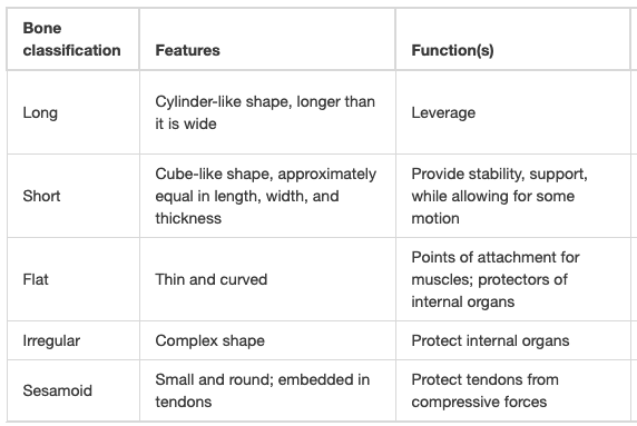
What do you think most of the classifications of bones are named after?
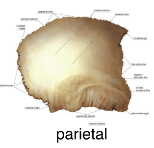
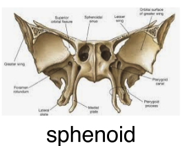
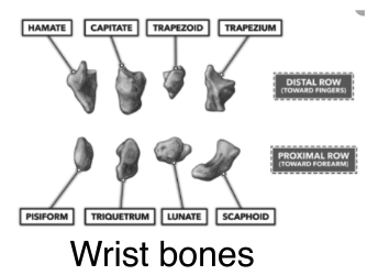
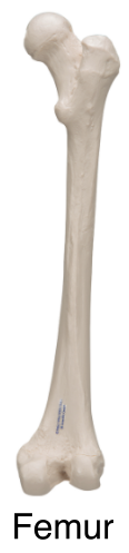
With your group, use the table above to match the bones with the correct type.
(I know that you may not know all of these bones yet - that's ok! Just look at the features in the table above and do your best.)
| Draggable item | arrow_right_alt | Corresponding Item |
|---|---|---|
(the lateral skull bones) | arrow_right_alt | Sesamoid |
(forms the base of your skull) | arrow_right_alt | Short |
| arrow_right_alt | Flat |
(your thigh bone) | arrow_right_alt | Irregular |
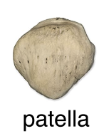
(your knee cap!) | arrow_right_alt | Long |
What are the structural and functional differences between the femur and the patella?
Examine the picture of the long bone below while you read the text below.
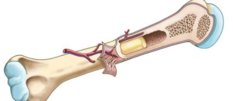
Long bones, like the femur and humerus (pictured above), consist of a elongated shaft (the diaphysis) and two enlarged ends (the epiphysis). Two types of bone tissue are found in the bone. The diaphysis is made primarily of compact bone, while the epiphysis are made of a lighter, "swiss cheese" looking bone called spongy bone. Where the epiphyses meet the diaphysis are the growth zones called the metaphysis. During the growing years, a plate of hyaline cartilage (the epiphyseal disk) exists in this area. These are commonly called the growth plates. As new cartilage forms on the epiphyseal side, it is destroyed and then replaced by bone on the diaphyseal side. The metaphysis, thus, consists of the epiphyseal disk, calcified cartilage, and bone during the growing years. At maturity, the area becomes completely ossified (hardened) and linear growth ceases.
Note that, in the central portion of the diaphysis, is a hollow chamber: the medullary cavity. The compact bone tissue of the shaft provides ample strength, so a solid cylinder of bone us unnecessary. This entire medullary cavity contains a fatty material called yellow marrow. The spongy bone of the epiphysis of the femur and humerus contain red marrow in the adult. The epiphyses of other long bones in the adult contain yellow marrow. Most red marrow in adults is found in the ribs, sternum, and vertebrae. Red marrow is the location of red blood cell production.
A tough covering, the periosteum, envelops the surface of the entire bone except for the areas of articulation. This covering consists of fibrous connective tissue which is quite vascular (has many blood vessels). The surfaces of each epiphysis which contact adjacent bones are covered with smooth articular cartilage which allows for smooth movement in joints.
Use the reading above to label the diagram below with the following terms (bolded above):
diaphysis, epiphysis, compact bone, epiphyseal disk, spongy bone, metaphysis, medullary cavity, yellow marrow, red marrow, periosteum, articular cartilage
Additionally, color the features listed in the 'key' with the required colors.
The matrix of bone is extremely hard due to the presence of mineral salts. These salts, calcium carbonate (CaCO3) and calcium phosphate (Ca3(PO4)2), are secreted by young bone cells called osteoblasts. Once ossification(hardening) is complete, the osteoblasts mature into osteocytes. Illustration A in the figure below shows a portion of the mandible enlarged to show the nature of bone tissue at different degrees of magnification. Two kinds of bone tissue, compact and spongy, can be seen in illustration B. Compact bone tissue is the dense outer portion. The inner part of the bone, which is sponge-like and porous, is the spongy bone. The bony processes between the open spaces in the spongy bone are called trabeculae.
Permeating the compact bone tissue is an intricate system of passageways, the Haversian canals. Haversian canals are small cavities in the bone that allow for nerves and blood vessels to carry nourishment throughout the bone to the living osteocytes ebmedded in the mineral salts. These canals are shown in the longitudinal cut face of the bone section in illustration B. Illustration C is an enlargement of an osteon - the term for a Haversian canal with a cluster of osteocytes arranged concentrically in the calcified bone matrix (also known as lamellae) around it. Illustration D shows an enlarged osteocyte. The tiny canals arranged around the osteocyte, called canaliculi, connect with adjacent cells. In this manner, all cells can get oxygen and nourishment from the Haversian canal.
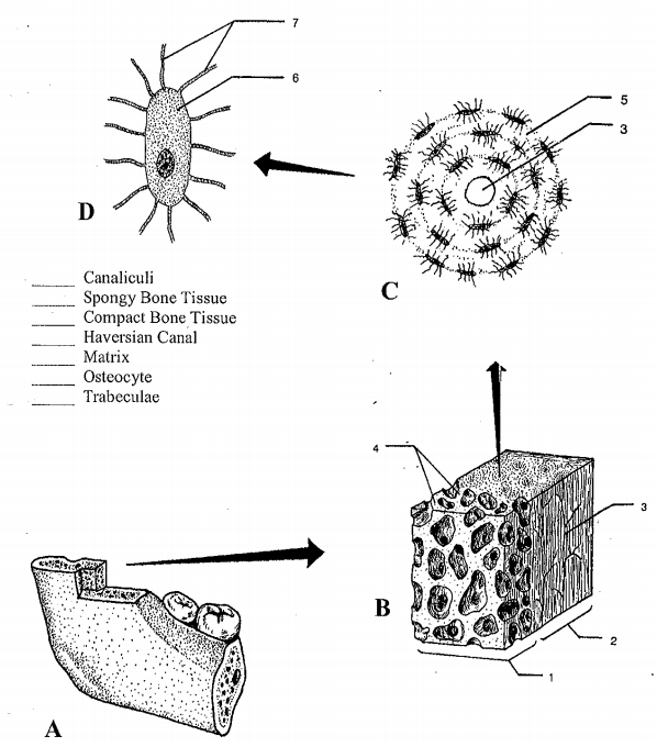
Using the reading and diagram above, match the numbers in the diagram with the correct term.
| Draggable item | arrow_right_alt | Corresponding Item |
|---|---|---|
5 | arrow_right_alt | spongy bone tissue |
3 | arrow_right_alt | compact bone tissue |
6 | arrow_right_alt | Haversian canal |
7 | arrow_right_alt | osteocyte |
2 | arrow_right_alt | trabeculae |
4 | arrow_right_alt | lamellae / matrix |
1 | arrow_right_alt | canaliculi |
PHEW! That's a lot of information and a LOT of terms -amiright?
Consider your comprehension of the terms from above. Categorize them below:
canaliculi
osteocyte
compact bone tissue
Haversian canal
spongy bone tissue
lamellae
trabeculae
I think I get this term!
I don't get this term quite yet
Ok, let's see if we can explain any of these better. Watch the two videos below, focusing on the terms presented above.
Feel free to change your answers to number 12 if need be!
Ok, let's try this again.
Consider your comprehension of the terms from above. Categorize them below (again!):
compact bone tissue
trabeculae
canaliculi
Haversian canal
spongy bone tissue
lamellae
osteocyte
I think I get this term!
I don't get this term quite yet
Ok, let's look at these things one more time to help you put it all together. Look for the terms from above in the diagram below. (I know this might feel repetative, but repetition is key in the world of A&P!)
Circle the terms in the diagram if they are present and add them if they are not:
spongy bone tissue
compact bone tissue
Haversian canal
osteocyte
trabeculae
lamellae
canaliculi
Ok, one last time (I promise!!)
Consider your comprehension of the terms from above. Categorize them below (again!):
spongy bone tissue
osteocyte
lamellae
Haversian canal
trabeculae
compact bone tissue
canaliculi
I think I get this term!
I don't get this term quite yet
Which of the following are components of an osteon?
Compare and contrast spongy bone and compact bone.
In your own words, compare and contrast the Haversian canal and canaliculi.
Draw a picture: What is one new thing that you found interesting from this lesson?
What is one question that you still have about today's lesson?