Copy of Skeletal Muscles: Sliding Filament Model (5/28/2026)
star
star
star
star
star
Last updated 3 months ago
17 Nsɛmmisa
1
1
1
1
1
1
2
1
1
2
5
8
2
2
4
2
5
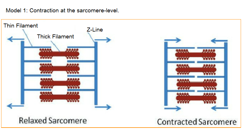
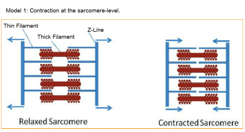
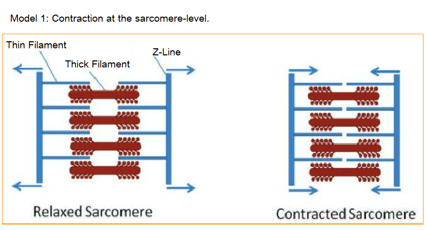
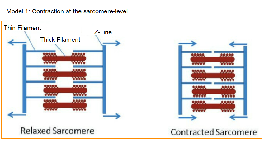
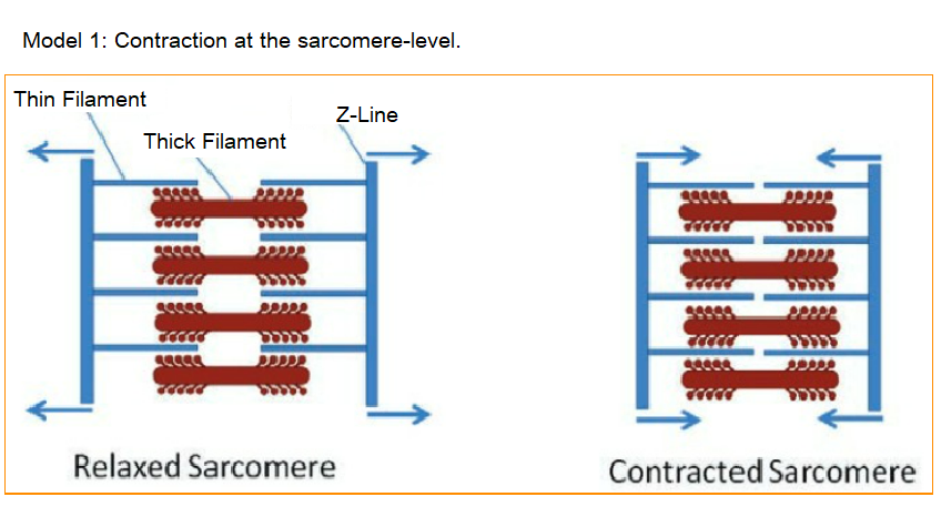
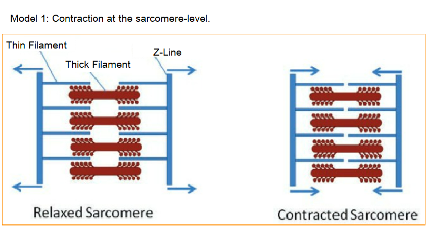
a.) What structural component of the sarcomere is associated with the arrows?
b.) Based on this, which component of the sarcomere is actually being moved when the sarcomere contracts and relaxes?
c.) Which component of the sarcomere is physically attached to the structure that gets moved (the answer to 'b')?
d.) Which component of the sarcomere is not directly attached to the Z-line?
e.) Based on your answer to 'd', what component of the sarcomere is likely pulling on the thin filaments to bring the Z-lines closer together?
Examine model 1 and your answers to 1a-e; in the next blanks, you will write a short description of the role of the thin filaments, thick filaments, and Z-line in sarcomere contraction.
Individual Work
Write a short description of the role of the Thin Filaments.
If you cannot figure it out, for half-credit, perform a web-search; but you must tell me in your answer that you did this.
Individual Work
Write a short description of the role of the Thick Filaments.
If you cannot figure it out, for half-credit, perform a web-search; but you must tell me in your answer that you did this.
Individual Work
Write a short description of the role of the Z-Lines.
If you cannot figure it out, for half-credit, perform a web-search; but you must tell me in your answer that you did this.
Click, "Show Your Work."
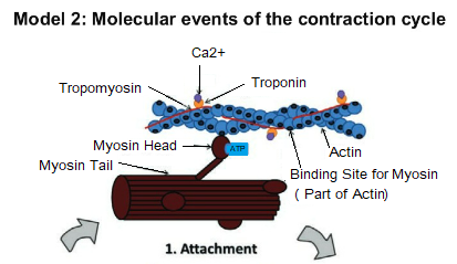
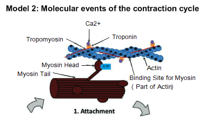
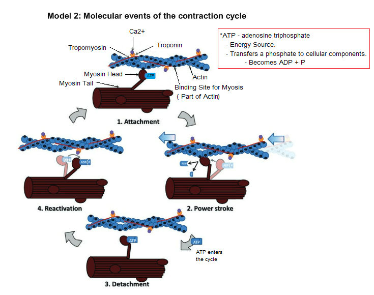
Find the thin filament in Part 1 (Attachment) of Model 2, circle it and label it, "thin filament."
You may have to zoom in (click the magnifying glass with the '+' sign, then click on the image).
To move the image around, click the hand, then click and drag the image.
Which component of the thin filament makes the main 'string of pearls' portion of the filament?
Individual Work
List the other components of the thin filament (I must re-grade this).
The myosin head is bound to two molecules; what are they?
To which specific region of the actin is the myosin head bound to?
Individual Work
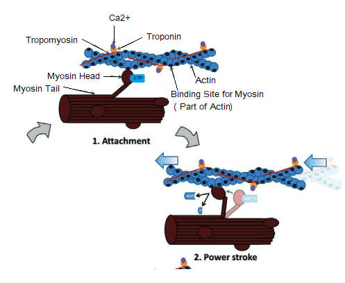
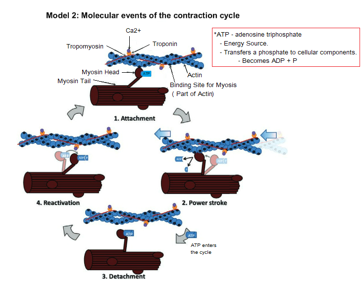
In your own words, describe the change that occurs in the myosin between stage 1 (attachment) and stage 2 (power stroke).
If you cannot figure it out, for half-credit, perform a web-search; but you must tell me in your answer that you did this.
a.) The myosin head doesn't release from the binding site on the actin until a new molecule attaches to the myosin head. According to stage 3 (detachment), what molecule allows the myosin to release from the binding site?
b.) In stage 4 of the cycle, the myosin head moves back into a cocked position. Where does it get the energy to re-cock itself?
c.) Look at the myosin binding sites on the actin filament. Does it appear that each site lines up perfectly with a myosin head?
d.) Myosin can only bind to a binding site that is at the tip of a helix in the actin filament (like the one in phase 1 of the model). After the power stroke (pulling phase) and detachment is the myosin head lined up with a binding site at the tip of the filament?
e.) Can this particular myosin head bind to the actin filament for the next power stroke?
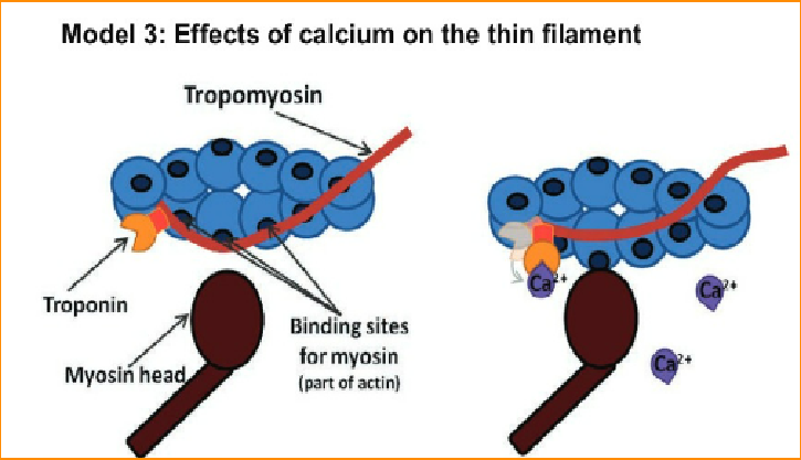
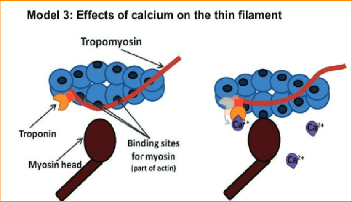
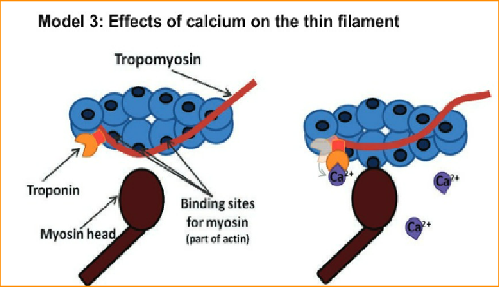
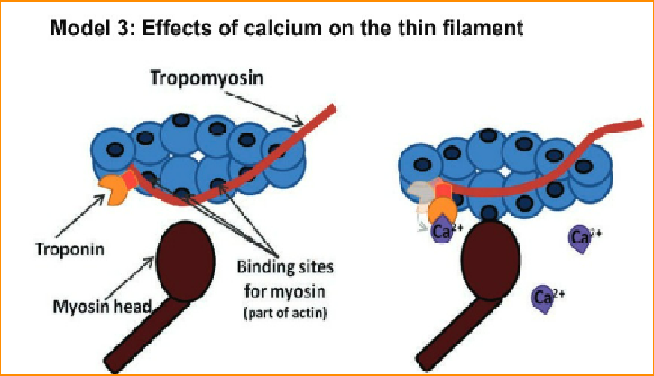
a.) Look at the left of model 3; is calcium present here?
b.) Is the myosin binding site of actin exposed so that myosin can actually bind?
c.) What is blocking myosin's access to the binding site of actin?
d.) Look at the right side of model 3; is calcium present here?
e.) To what molecule did it bind to?
f.) What changed about the troponin after it bound to calcium?
g.) What effect did this change in troponin have on tropomyosin?
h.) Is the myosin binding site exposed when calcium is bound to troponin?
Individual Work
Write a sentence that explains the role of troponin, tropomyosin, and calcium in muscle cell contraction.
If you cannot figure it out, for half-credit, perform a web-search; but you must tell me in your answer that you did this.
Individual Work
What might happen to a muscle cell in either of the following situations?
a.) The cell runs out of ATP.
b.) There is an excess amount of calcium in the cell.
If you cannot figure it out, for half-credit, perform a web-search; but you must tell me in your answer that you did this.
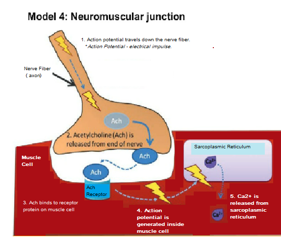
a.) Can muscles contract if there isn't any free calcium in the cell?
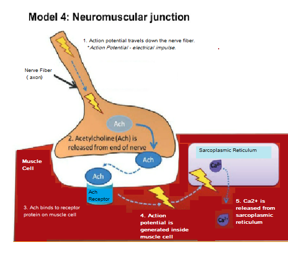
b.) According to model 4, where is calcium stored in a muscle cell?
c.) What causes calcium to be released from where it is stored?
d.) What chemical messenger transmits the electrical impulse between the nerve cell and the muscle cell (use the actual name, not the abbreviation)?
Individual Work
Acetylcholine esterase is an enzyme that removes acetylcholine from the neuromuscular junction. The nerve gas sarin blocks this enzyme causing acetylcholine to remain bound to the muscle cell receptors.
If acetylcholine stays bound to its receptor, how will that affect calcium levels inside the cell? Would the cell be able to moderate whether or not it contracts or relaxes (would it be stuck in either a contracted or relaxed state)?
If you cannot figure it out, for half-credit, perform a web-search; but you must tell me in your answer that you did this.
Individual Work
Use all four models to put the following events in order from the signal of the brain reaching a muscle to the contraction of the whole muscle.
If you cannot figure it out, for half-credit, perform a web-search; but you must tell me in your answer that you did this.
The myofibril (muscle fiber) gets shorter (contracts)
Acetylcholine binds to receptors on the muscle cell membrane and causes the electrical impulse to be transmitted to the muscle cell
The nerve impulse reaches the end of the nerve and causes it to release acetylcholine (Ach)
The myosin head binds the myosin binding domain of actin
Myosin bends in two places, releasing ADP and pulling on the thin filament
Rotation of troponin move tropomyosin off the myosin binding site on actin
The Z-lines are pulled closer together and the A-band shrinks
The electrical impulse inside the muscle cell causes the release of calcium ions from the sarcoplasmic reticulum
Calcium ions bind to troponin causing it to rotate
An electrical impulse travels down a nerve fiber