Use the reading and any diagrams on the left-hand side to complete the reading guide on the right-hand side.
Each section of the reading will also have an audio recording of it being read attached just above the section of reading that it corresponds to. However, if you use this, you should follow along with the text as the audio plays.
There are three sections to this activity — 1.) Chromosomes; 2.) Mitosis and Cytokinesis; 3.) Exit Ticket.
When complete with the first and second sections, complete the exit ticket and then submit this.
Exit Ticket - self-reflective; when you are asked about your confidence regarding this concept, it is not asking if you understand everything or even most of it. Instead, consider how many new things you were introduced to or learned and then ask if you will be able to learn if given the opportunity and the time.
Use the reading and any diagrams on the left-hand side to complete the reading guide on the right-hand side.
Each section of the reading will also have an audio recording of it being read attached just above the section of reading that it corresponds to. However, if you use this, you should follow along with the text as the audio plays.
There are three sections to this activity — 1.) Chromosomes; 2.) Mitosis and Cytokinesis; 3.) Exit Ticket.
When complete with the first and second sections, complete the exit ticket and then submit this.
Exit Ticket - self-reflective; when you are asked about your confidence regarding this concept, it is not asking if you understand everything or even most of it. Instead, consider how many new things you were introduced to or learned and then ask if you will be able to learn if given the opportunity and the time.
B.4.4 Use a model to illustrate the role of cellular division (mitosis) and differentiation in producing and maintaining complex organisms.
Chromosomes and their structure
How genes are related to chromosomes
The number of chromosomes in human cells
During cell division in eukaryotic cells, the nucleus divides before the cell itself divides. The process in which the nucleus divides is called mitosis. Before mitosis occurs, a cell’s DNA is replicated. This is necessary so that each daughter cell will have the correct amount of DNA and the correct type of chromosomes from the parent cell. How is the replicated DNA sorted and separated so that each daughter cell gets a complete set of the genetic material? To understand how this happens, you need to know more about chromosomes.
Chromosomes are coiled structures made of DNA wrapped around histone (a type of protein). Chromosomes are the form of the genetic material of a cell during cell division. It is this coiled structure that ensures the proper segregation (distribution) of the chromosomes during cell division. During other phases of the cell cycle, DNA is not coiled into chromosomes. Instead, it exists as a grainy material called chromatin. The primary difference between a chromosome and chromatin has to do with how condensed the DNA is, or how tightly coiled it is around the histone proteins. Chromosomes are more condensed (more tightly wrapped around histone) than chromatin is.
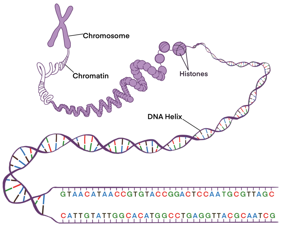
In eukaryotic cells, the
Chromosomes are made out of two structures -
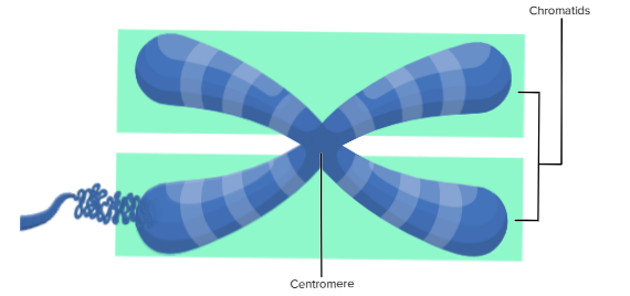
DNA condenses and coils into the familiar X-shaped form of a chromosome only after it has replicated. Because DNA has already replicated prior to cell division, each chromosome actually consists of two identical copies. The two copies are called sister chromatids. They are attached to one another at a region called the centromere.
The chromosomes in human cells normally have two sets of chromosomes for each type of chromosome, one set (type) is inherited from each parent. In other words, each chromosome type is in a pair. There are 23 chromosomes in each set, for a total of 46 chromosomes per cell. Each chromosome in one set is matched by a chromosome of the same type in the other set, so there are actually 23 pairs of chromosomes per cell. Each pair consists of chromosomes of similar size and shape (that can be used to pair them up) that also contain the same genes. The chromosomes in a pair are known as homologous chromosomes.
Prior to cell division, the DNA
In humans, each chromosome type is in
B.4.4 Use a model to illustrate the role of cellular division (mitosis) and differentiation in producing and maintaining complex organisms.
Learning Goals:
The process of cell division in eukaryotic cells through mitosis.
The four stages of mitosis.
How cytokinesis occurs differently in plant and animal cells.
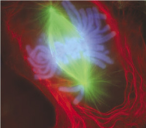
What do you think this colorful picture shows? If you guessed that it’s a picture of a cell undergoing cell division, you are right. But more specifically, the image is a lung cell stained with fluorescent dyes undergoing mitosis, during early anaphase.
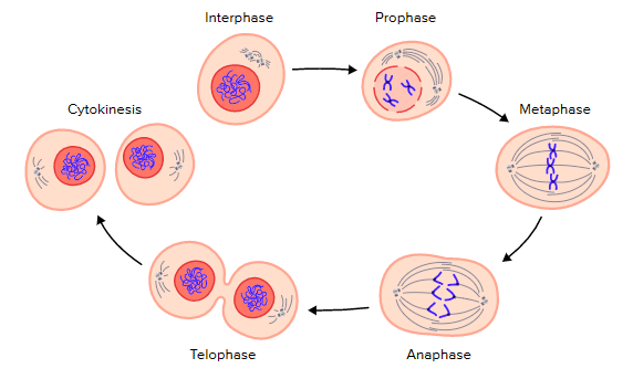
During mitosis, the division of the nucleus in a eukaryotic cell occurs in four stages — prophase, metaphase, anaphase, and telophase. These stages, along with the main cellular structures observed during mitosis, are shown below and are described in greater detail in the following sections. Mitosis occurs just prior to, and works in conjunction with, cytokinesis, which is the division of the cytoplasm and the rest of the cell.
Mitosis is the division of the
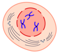
The first and longest phase of mitosis is prophase (Figure below). During prophase, chromatin condenses into chromosomes, and the nuclear envelope, or membrane, breaks down. In animal cells, the centrioles near the nucleus begin to separate and move to opposite poles (sides) of the cell. As the centrioles move, a spindle starts to form between them. The spindle consists of fibers made of microtubules that are used by motor proteins to pull chromosomes apart during cell division.
Prophase: DNA condenses to form visible chromosomes, nuclear membrane breaks down, spindle fibers begin to form.
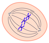
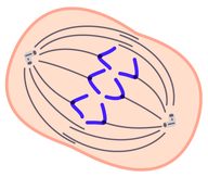
During metaphase, spindle fibers attach to the centromere of each pair of sister chromatids (Figure below). The sister chromatids line up at the equator, or center, of the cell. This is also known as the metaphase plate. The spindle fibers along with motor proteins on the centromere ensure that the sister chromatids of each chromosome will separate and go to different daughter cells when the cell divides.
Metaphase: chromosomes line up at the center of the cell; spindle fibers attach to both sides of the centromere; chromosomes are still made up of sister chromatids.
The first two stages of mitosis are
Prophase:
DNA
Nuclear membrane
Metaphase:
Chromosomes
Spindle fibers attach to
During anaphase (Figure below), sister chromatids separate and move to opposite sides of the cell. The sister chromatids are pulled apart by motor proteins located on the centromere moving down each spindle fiber. As the motor protein moves down the spindle fiber, the spindle fibers begin to break down providing the illusion that the spindle fibers are pulling on the chromatids, but this is not the case. One sister chromatid moves to one pole of the cell, and the other sister chromatid moves to the opposite pole. At the end of anaphase, each pole of the cell has a complete set of chromosomes.
Anaphase: sister chromatids are pulled apart and move to opposite poles of the cell.
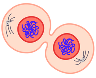
During telophase (Figure below), the chromosomes begin to uncoil and form chromatin. This prepares the genetic material for directing the metabolic activities of the new cells. The spindle also breaks down, and new nuclear membranes (nuclear envelope) form.
Telophase: spindle fibers break down, two nuclear membranes form, and chromosomes begin to uncondense to form chromatin; cytokinesis overlaps at the end of telophase.
The last two stages of mitosis are
Anaphase:
Sister chromatids are
Telophase:
Spindle fibers
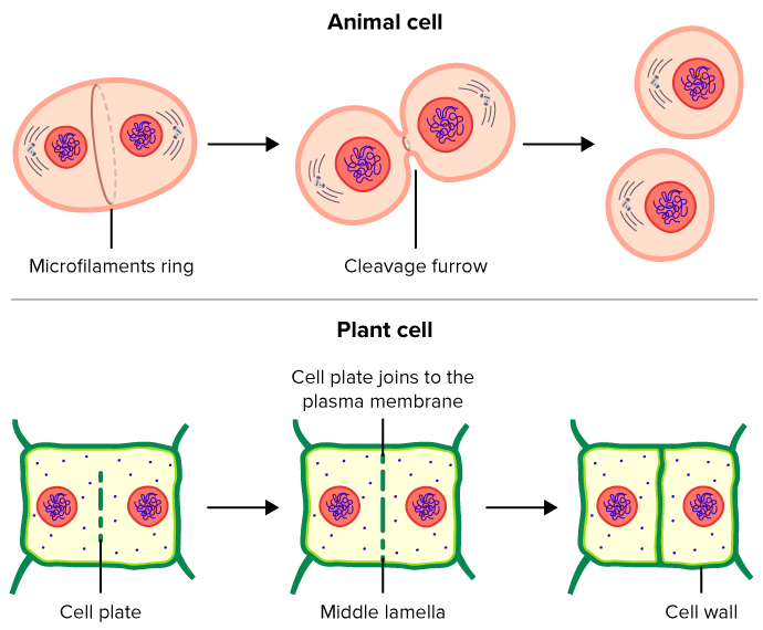
Cytokinesis is the final stage of cell division in eukaryotes as well as in prokaryotes. During cytokinesis, the cytoplasm splits in two and the cell divides resulting in two daughter cells. Cytokinesis occurs somewhat differently in plant and animal cells, shown below. In animal cells, the cell membrane of the parent cell pinches inward along the cell’s equator (the middle of the cell) until two daughter cells form. In plant cells, a cell plate forms along the equator of the parent cell. The cell plate will become a cell wall. Finally, a new cell membrane will form along each side of the cell plate.
Cytokinesis will divide the
In plant and animal cells, cytokinesis operates differently. In animal cells, the
Provide a list of one or two things that you learned or confusions that were cleared up from this activity.
After having completed this activity, my confidence (not your intellectual mastery) regarding this concept has: