Log in
Sign up for FREE
arrow_back
Library

Copy of 2.) Mitosis and Cell Cycle Models (5/28/2026)

star
star
star
star
star
Last updated about 2 hours ago
23 questions
Mitosis and Cell Cycle
1
Exit Ticket
Required
0
Required
0
4
2
2
1
2
1
Read This:

A chromosome is a structure made mainly of two components:
  • DNA
  • Proteins called histones
The DNA wraps around the histones, which helps keep it organized, similar to how folding clothes keeps them neat, and allows it to fit inside a cell (in prokaryotes) or inside the nucleus (in eukaryotes).

When a cell is preparing to divide (through mitosis or meiosis, followed by cytokinesis), the DNA wraps more tightly around the histones than usual. This tighter wrapping causes the chromosomes to condense, meaning they become more compact and take up less space. Because of this, chromosomes become visible under a light microscope at this stage. At other times, they are not easily seen and cannot be clearly distinguished from the rest of the nucleus.
Model 2 - The Cell Cycle:
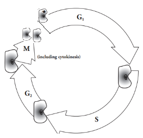

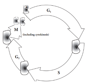
In the model of the cell cycle, it begins at the flat part of the G1 arrow with the one cell with the asterisk (*) and ends at the tip of the M arrow with two cells, both without an asterisk.

Notice what happens to the size of the cell from the beginning of G1 to end of G1 (near the beginning of S).
1
1
1
1
4
1
1
1
1
Question 16
16.


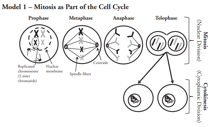
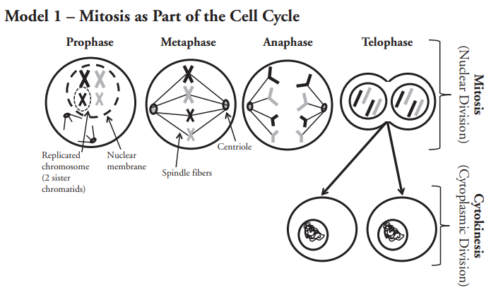
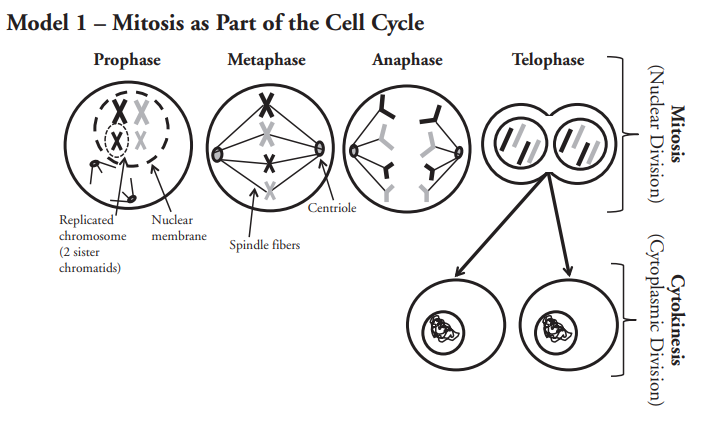
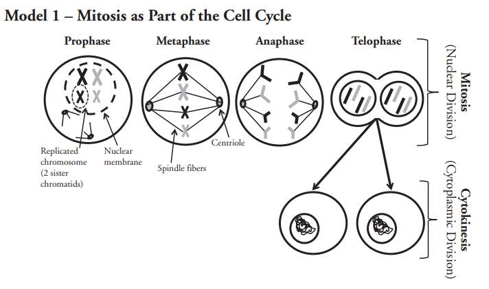
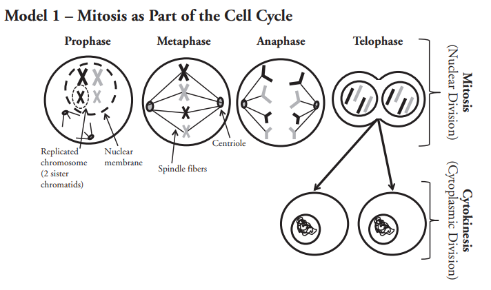
Cellular division has two parts — mitosis (the division of the nucleus) and cytokinesis (the division of the cytoplasm and the rest of the cell) — that will form two new daughter cells. Using model 1 and the cell cycle model, above, and what you have learned thus far, provide a logical reason why mitosis needs to come before cytokinesis during the cell division.

1
1
1
1
1
Question 22
22.

After having completed this activity, provide a list of one or two things that you have learned or anything that you understand better than you did prior to this activity.

Question 23
23.


Model 2 - The Cell Cycle:


Read This:

In the model of the cell cycle, it begins at the flat part of the G1 arrow with the one cell with the asterisk (*) and ends at the tip of the M arrow with two cells, both without an asterisk.

Notice what happens to the size of the cell from the beginning of G1 to end of G1 (near the beginning of S).
Question 1
1.
Using model 1, list the four steps of mitosis in order.
1st _______
2nd _______
3rd _______
4th _______
Question 2
2.
Where is mitosis in the cell cycle? It occurs before, or prior to, _______ and after _______.
Question 3
3.
Refer to the cell cycle shown at the left.

a.) How many cells are present at the begining of mitosis?
_______

b.) How many cells are present at the end of mitosis?
_______
Question 4
4.
Refer back to model 1. Look for the replicated chromosomes in the cell in prophase.

How many chromatids are present in each replicated chromosome? _______
Question 5
5.
How many replicated chromosomes are in the original cell in prophase of model 1 (they look like X's)?
_______

How many single chromosomes are in each new cell in telophase (they look like I's)?
_______
Question 6
6.
Did the number of chromosomes (not chromatids) change from prophase to the end of telophase?
_______
Question 7
7.

Describe what each new cell at the end of telophase would contain, with respect to chromosome number, if replication of chromosomes (DNA) did not occur prior to mitosis.

Question 8
8.

If the situation in the previous question occurred, would these new cells be able to survive? Why do you think this?

Question 9
9.

The S phase of the cell cycle stands for synthesis, which means to make or build something more complex out of simpler parts. Scientists know that during the S phase DNA is being made in the nucleus of the cell. Why do you think the cell needs to make more DNA at this time in the cell cycle?

Question 10
10.
Refer to Model 1.

The chromosomes that are shaped like “X” (made of two sister chromatids) have double the amount of DNA than the chromosomes that are shaped like “I.”

During what phase of the cell cycle do you think the chromosomes are replicated (copied)?
_______
Question 11
11.
Refer to Model 1.

a. In which phase of mitosis do you see the spindle fibers forming? _______

b. At what phase of mitosis do the replicated chromosomes (sister chromatids) separate? _______

c. In which phase do you see that the spindle fibers have disappeared? _______

d. Look at metaphase and anaphase. Suggest the purpose of the spindle fibers during mitosis. _______
Question 12
12.
Refer to Model 1.
What happens to the nuclear membrane during and after prophase. _______
Question 13
13.

Why might it be imporant for the nuclear envelope to breakdown during mitosis?

Question 14
14.
At what point during mitosis has the nuclear membrane reformed? _______
Question 15
15.
What is actually dividing during cytokinesis? _______
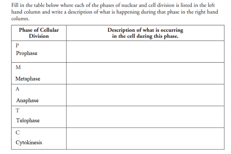
For 17 - 21, refer to Model 1 to help you describe the basic events of each phase cell division.

In the table to the bottom-left, each blank has a number in a circle, these correspond to the numbered questions to the right of the table.

Use your note-taking sheet, but do not look things up on the internet - I can tell when you do this.

Try to restrict your comments to what the nucleus and chromosomes are doing. In other words, be brief and get to the point.
Question 17
17.

Describe the primary events of prophase.

Question 18
18.

Describe the primary events of metaphase.

Question 19
19.

Describe the primary events of anaphase.

Question 20
20.

Describe the primary events of telophase.

Question 21
21.

Describe the primary events of cytokinesis.

After having completed this activity, my confidence regarding this concept has:
Significantly increased
Moderately increased
Somewhat increased
Not changed
Somewhat decreased
Moderately decreased
Significantly increased