Copy of Meiosis Reading Guide - OpenStax (5/28/2026)
star
star
star
star
star
Last updated about 2 hours ago
0 questions
Meiosis and Sexual Reproduction
Sexual reproduction requires the union of two specialized cells, called gametes, each of which contains one set of chromosomes. When gametes unite, they form a zygote, or fertilized egg that contains two sets of chromosomes. (Note: Cells that contain one set of chromosomes are called haploid; cells containing two sets of chromosomes are called diploid.) If the reproductive cycle is to continue for any sexually reproducing species, then the diploid cell must somehow reduce its number of chromosome sets to produce haploid gametes; otherwise, the number of chromosome sets will double with every future round of fertilization. Therefore, sexual reproduction requires a nuclear division that reduces the number of chromosome sets by half.
Most animals and plants and many unicellular organisms are diploid and therefore have two sets of chromosomes. In each somatic cell of the organism (all cells of a multicellular organism except the gametes or reproductive cells), the nucleus contains two copies of each chromosome, called homologous chromosomes. Homologous chromosomes are matched pairs containing the same genes in identical locations along their lengths. Diploid organisms inherit one copy of each homologous chromosome from each genetic contributor.
Meiosis is the nuclear division that forms haploid cells from diploid cells, and it employs many of the same cellular mechanisms as mitosis. However, as you have learned, mitosis produces daughter cells whose nuclei are genetically identical to the original parent nucleus. In mitosis, both the parent and the daughter nuclei are at the same “ploidy level”—diploid in the case of most multicellular animals. Plants use mitosis to grow as sporophytes, and to grow and produce eggs and sperm as gametophytes; so they use mitosis for both haploid and diploid cells (as well as for all other ploidies). In meiosis, the starting nucleus is always diploid and the daughter nuclei that result are haploid. To achieve this reduction in chromosome number, meiosis consists of one round of chromosome replication followed by two rounds of nuclear division. Because many events that occur during each of the division stages are analogous to the events of mitosis, the same stage names are assigned. However, because there are two rounds of division, the major process and the stages are designated with a “I” or a “II.” Thus, meiosis I is the first round of meiotic division and consists of prophase I, prometaphase I, and so on. Likewise, Meiosis II (during which the second round of meiotic division takes place) includes prophase II, prometaphase II, and so on.
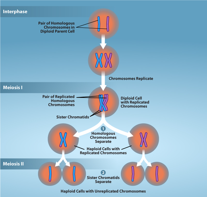
Figure 11.2 Overview of Meiosis. The production of gametes is a crucial process for sexually reproducing organisms. Meiosis is the mechanism used to reduce diploid cells to haploid gametes while introducing genetic diversity. Prior to meiosis, chromosomes are replicated in S-phase to ensure proper number of chromosomes in the resulting gametes. During meiosis, two successive rounds of division reduces the number of chromosomes (ploidy) of the cell by half. going from diploid cells to haploid gametes.
Meiosis I
Meiosis is preceded by an interphase consisting of G1, S, and G2 phases, which are nearly identical to the phases preceding mitosis. The G1 phase (the “first gap phase”) is focused on cell growth. During the S phase—the second phase of interphase—the cell copies or replicates the DNA of the chromosomes. Finally, in the G2 phase (the “second gap phase”) the cell undergoes the final preparations for meiosis.
During DNA duplication in the S phase, each chromosome is replicated to produce two identical copies—sister chromatids that are held together at the centromere by cohesin proteins, which hold the chromatids together until anaphase II. (Note: these chromosome copies are called sister chromatids regardless of whether they are in a female gamete or a male gamete.)
There are four stages that occur during meiosis I - prophase I, metaphase I, anaphase I, and telophase I.
Prophase I
Early in prophase I, before the chromosomes can be seen clearly with a microscope, the homologous chromosomes are attached at their tips to the nuclear envelope by proteins. As the nuclear envelope begins to break down, the proteins associated with homologous chromosomes bring the pair closer together. Recall that in mitosis, homologous chromosomes do not pair together. The synaptonemal complex, a lattice of proteins between the homologous chromosomes, first forms at specific locations and then spreads outward to cover the entire length of the chromosomes. The tight pairing of the homologous chromosomes is called synapsis. In synapsis, the genes on the chromatids of the homologous chromosomes are aligned precisely with each other. The synaptonemal complex supports the exchange of chromosomal segments between homologous nonsister chromatids—a process called crossing over. Crossing over can be observed visually after the exchange as chiasmata (singular = chiasma) (Figure 11.3).
In humans, even though the X and Y sex chromosomes are not completely homologous (that is, most of their genes differ), there is a small region of homology that allows the X and Y chromosomes to pair up during prophase I. A partial synaptonemal complex develops only between the regions of homology.
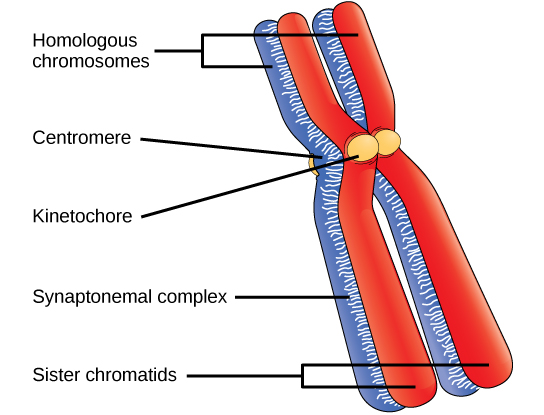
Figure 11.3 Early in prophase I, homologous chromosomes come together to form a synapse. The chromosomes are bound tightly together and in perfect alignment by a protein lattice called a synaptonemal complex and by cohesin proteins at the centromere.
Located at intervals along the synaptonemal complex are large protein assemblies called recombination nodules. These assemblies mark the points of later chiasmata and mediate the multistep process of crossover—or genetic recombination—between the nonsister chromatids. Near the recombination nodule, the double-stranded DNA of each chromatid is cleaved, the cut ends are modified, and a new connection is made between the nonsister chromatids. As prophase I progresses, the synaptonemal complex begins to break down and the chromosomes begin to condense. When the synaptonemal complex is gone, the homologous chromosomes remain attached to each other at the centromere and at chiasmata. The chiasmata remain until anaphase I. The number of chiasmata varies according to the species and the length of the chromosome. There must be at least one chiasma per chromosome for proper separation of homologous chromosomes during meiosis I, but there may be as many as 25. Following crossover, the synaptonemal complex breaks down and the cohesin connection between homologous pairs is removed. At the end of prophase I, the pairs are held together only at the chiasmata (Figure 11.4). These pairs are called tetrads because a total of four sister chromatids of each pair of homologous chromosomes are now visible.
The crossover events are the first source of genetic variation in the nuclei produced by meiosis. A single crossover event between homologous nonsister chromatids leads to a reciprocal exchange of equivalent DNA between an egg-derived chromosome and a sperm-derived chromosome. When a recombinant sister chromatid is moved into a gamete cell it will carry a combination of maternal and paternal genes that did not exist before the crossover. Crossover events can occur almost anywhere along the length of the synapsed chromosomes. Different cells undergoing meiosis will therefore produce different recombinant chromatids, with varying combinations of maternal and parental genes. Multiple crossovers in an arm of the chromosome have the same effect, exchanging segments of DNA to produce genetically recombined chromosomes.